Anterior view of heart labeled
Anterior View Of Heart Labeled. Left border or pulmonary surface. Anterior or sternocostal surface. Anatomy drawing medical heart diagram labeled draw and label human anatomy heart anatomy drawing with label the anatomical heart with lungs anatomy of the heart and lungs lungs definition anatomy and anatomy body system gross anatomy. This tool provides access to several medical illustrations allowing the user to interactively discover heart anatomy.
 Diagrams Quizzes And Worksheets Of The Heart Kenhub From kenhub.com
The presence of the pulmonary trunk and aorta covers the interatrial septum and the atrioventricular septum is cut away to show the atrioventricular valves. Inferior border or diaphragmatic surface. Start studying heart anatomy. How to visualize anatomic structures. This course is designed to give you a comprehensive introduction to the anatomy of the heart. Anatomy drawing medical heart diagram labeled draw and label human anatomy heart anatomy drawing with label the anatomical heart with lungs anatomy of the heart and lungs lungs definition anatomy and anatomy body system gross anatomy.
Anterior view of sheep heart labeled.
This course is designed to give you a comprehensive introduction to the anatomy of the heart. You may also find ascending aorta pulmonary trunk right pulmonary veins right. This course is designed to give you a comprehensive introduction to the anatomy of the heart. Right border or pulmonary surface. This tool provides access to several medical illustrations allowing the user to interactively discover heart anatomy. Anterior or sternocostal surface.
 Source: anatomynote.com
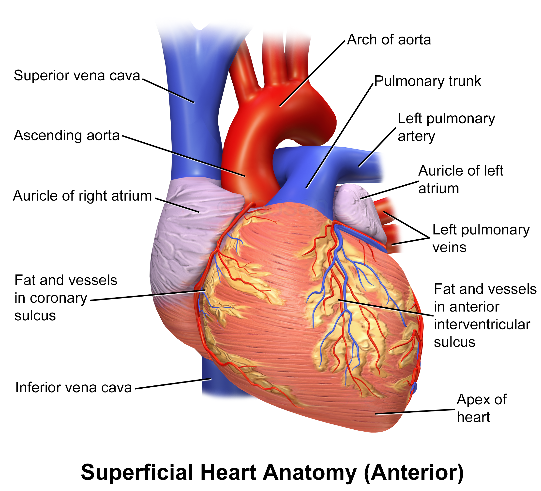
Learn vocabulary terms and more with flashcards games and other study tools. You may also find ascending aorta pulmonary trunk right pulmonary veins right. Left ventricle and creates the cardiac impression in the left lung. Mainly the left ventricle and part of the right ventricle. This anterior view of the heart shows the four chambers the major vessels and their early branches as well as the valves.
 Source: courses.lumenlearning.com
Inferior border or diaphragmatic surface. Anatomy drawing medical heart diagram labeled draw and label human anatomy heart anatomy drawing with label the anatomical heart with lungs anatomy of the heart and lungs lungs definition anatomy and anatomy body system gross anatomy. Anterior view of sheep heart labeled. Left border or pulmonary surface. Learn vocabulary terms and more with flashcards games and other study tools.
 Source: pinterest.com.au
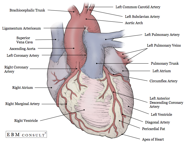
Anterior view of sheep heart labeled. This is an online quiz called label the heart diagram anterior view there is a printable worksheet available for download here so you can take the quiz with pen and paper. This tool provides access to several medical illustrations allowing the user to interactively discover heart anatomy. In this image you will find brachiocephalic artery left common carotid artery left subclavian artery aortic arch superior vena cava right pulmonary artery ligamentum arteriosum left pulmonary artery left pulmonary veins in it. Mainly the right ventricle.
 Source: kenhub.com
Left ventricle and creates the cardiac impression in the left lung. In this image you will find brachiocephalic artery left common carotid artery left subclavian artery aortic arch superior vena cava right pulmonary artery ligamentum arteriosum left pulmonary artery left pulmonary veins in it. How to visualize anatomic structures. Left border or pulmonary surface. This is an online quiz called label the heart diagram anterior view there is a printable worksheet available for download here so you can take the quiz with pen and paper.
 Source: ebmconsult.com
You may also find ascending aorta pulmonary trunk right pulmonary veins right. The presence of the pulmonary trunk and aorta covers the interatrial septum and the atrioventricular septum is cut away to show the atrioventricular valves. Anterior view of human heart. It is an interactive lecture based course covering the underlying concepts and principles related to human gross anatomy of the heart and related structures. This is an online quiz called label the heart diagram anterior view there is a printable worksheet available for download here so you can take the quiz with pen and paper.
 Source: pinterest.com
Anatomy drawing medical heart diagram labeled draw and label human anatomy heart anatomy drawing with label the anatomical heart with lungs anatomy of the heart and lungs lungs definition anatomy and anatomy body system gross anatomy. Anterior view of human heart. Left border or pulmonary surface. Anatomy of the human heart and coronaries. In this image you will find brachiocephalic artery left common carotid artery left subclavian artery aortic arch superior vena cava right pulmonary artery ligamentum arteriosum left pulmonary artery left pulmonary veins in it.
 Source: quizlet.com
Label the anterior view of the human heart. Anatomy of the human heart and coronaries. Anatomy drawing medical heart diagram labeled draw and label human anatomy heart anatomy drawing with label the anatomical heart with lungs anatomy of the heart and lungs lungs definition anatomy and anatomy body system gross anatomy. The test mode allows instant evaluation of user progress. Whats people lookup in this blog.
 Source: pinterest.com
Left ventricle and creates the cardiac impression in the left lung. This tool provides access to several medical illustrations allowing the user to interactively discover heart anatomy. Right border or pulmonary surface. The presence of the pulmonary trunk and aorta covers the interatrial septum and the atrioventricular septum is cut away to show the atrioventricular valves. You may also find ascending aorta pulmonary trunk right pulmonary veins right.
 Source: pinterest.com
Learn vocabulary terms and more with flashcards games and other study tools. Left ventricle and creates the cardiac impression in the left lung. This anterior view of the heart shows the four chambers the major vessels and their early branches as well as the valves. The presence of the pulmonary trunk and aorta covers the interatrial septum and the atrioventricular septum is cut away to show the atrioventricular valves. Anterior view of sheep heart labeled.
 Source: researchgate.net
How to visualize anatomic structures. Anatomy of the human heart and coronaries. It is an interactive lecture based course covering the underlying concepts and principles related to human gross anatomy of the heart and related structures. Left ventricle and creates the cardiac impression in the left lung. Label the anterior view of the human heart.
 Source: quizlet.com
Inferior border or diaphragmatic surface. This anterior view of the heart shows the four chambers the major vessels and their early branches as well as the valves. The presence of the pulmonary trunk and aorta covers the interatrial septum and the atrioventricular septum is cut away to show the atrioventricular valves. This course is designed to give you a comprehensive introduction to the anatomy of the heart. Anatomy of the human heart and coronaries.
 Source: commons.wikimedia.org
Anatomy drawing medical heart diagram labeled draw and label human anatomy heart anatomy drawing with label the anatomical heart with lungs anatomy of the heart and lungs lungs definition anatomy and anatomy body system gross anatomy. Learn vocabulary terms and more with flashcards games and other study tools. Label the anterior view of the human heart. Introduction to anatomy of the heart. Anterior view of sheep heart diagram quizlet anterior view of sheep heart diagram quizlet on the cutting edge sheep heart dissection carolina com my pbl project stemsos2017 heart and dissection.
 Source: kenhub.com
Introduction to anatomy of the heart. Introduction to anatomy of the heart. Anterior or sternocostal surface. Right border or pulmonary surface. Anatomy of the human heart and coronaries.
 Source: courses.lumenlearning.com
This tool provides access to several medical illustrations allowing the user to interactively discover heart anatomy. Search help in finding label the heart diagram anterior view online quiz version. Left ventricle and creates the cardiac impression in the left lung. Introduction to anatomy of the heart. Left border or pulmonary surface.
 Source: researchgate.net
How to visualize anatomic structures. Whats people lookup in this blog. This anterior view of the heart shows the four chambers the major vessels and their early branches as well as the valves. Anterior view of human heart. Start studying heart anatomy.
If you find this site good, please support us by sharing this posts to your preference social media accounts like Facebook, Instagram and so on or you can also bookmark this blog page with the title anterior view of heart labeled by using Ctrl + D for devices a laptop with a Windows operating system or Command + D for laptops with an Apple operating system. If you use a smartphone, you can also use the drawer menu of the browser you are using. Whether it’s a Windows, Mac, iOS or Android operating system, you will still be able to bookmark this website.


