Bacteria microscope magnification
Bacteria Microscope Magnification. Why bacteria are difficult to see. Another bonus for this microscope is that it can be plugged in or powered by aa batteries when on the go. The magnification of microscopes comes from the ability of convex lenses to bend and focus light rays. Coli under the microscope at 400x.
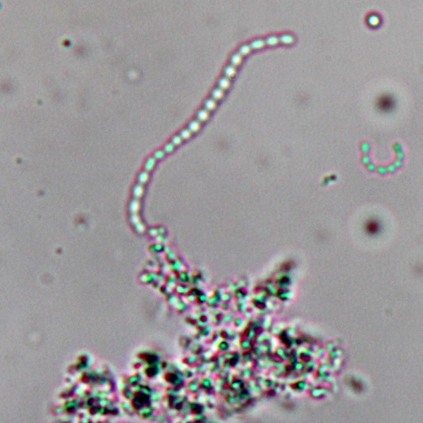
 Observing Bacteria Under The Light Microscope Microbehunter Microscopy From microbehunter.com
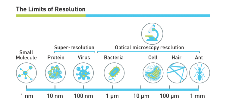
This bacteria is known as hay bacillus or grass bacillus. Whereas vibrio bacteria appear comma shaped spirilla are the type that appear spiral in shape. It is enough to see things as small as red blood cells 8 µm and bacteria 1 µm. In order to see bacteria you will need to view them under the magnification of a microscopes as bacteria are too small to be observed by the naked eye. While some people may confuse the two when viewed under the microscope they are different when students compare them under high magnification. From deep within the soil to inside the digestive tract of humans.
It is enough to see things as small as red blood cells 8 µm and bacteria 1 µm.
This bacteria is known as hay bacillus or grass bacillus. The amscope 40x 1000x compound microscope is a solid affordable choice for a compound microscope for students and at home stem exploration. Light microscopy has been traditionally used for identifying bacteria but is often limited by inadequate resolution. Whereas vibrio bacteria appear comma shaped spirilla are the type that appear spiral in shape. Coli escherichia coli is a gram negative rod shaped bacterium. The optics must be good in order to resolve them properly at this magnification.
 Source: westlab.com.au
Bacteria are difficult to see with a bright field compound microscope for several reasons. In order to see their shape it is necessary to use a magnification of about 400x to 1000x. Whereas vibrio bacteria appear comma shaped spirilla are the type that appear spiral in shape. The prepared microscope slide image of bacillus subtilis at left was captured at 400x magnification. From deep within the soil to inside the digestive tract of humans.
 Source: microbehunter.com
While some people may confuse the two when viewed under the microscope they are different when students compare them under high magnification. In the 16 th century dutch microbiologist antonie van leeuwenhoek was first to see bacteria under a microscope. From deep within the soil to inside the digestive tract of humans. In order to see their shape it is necessary to use a magnification of about 400x to 1000x. The optics must be good in order to resolve them properly at this magnification.
Source: quora.com
Whereas vibrio bacteria appear comma shaped spirilla are the type that appear spiral in shape. Upon viewing the bacteria under the microscope you will be able to identify the bacteria based on a wide variety of physical characteristics. Most bacteria are 0 2 um in diameter and 2 8 um in length with a number of shapes ranging from spheres to rods and spirals. The amscope 40x 1000x compound microscope is a solid affordable choice for a compound microscope for students and at home stem exploration. In the 16 th century dutch microbiologist antonie van leeuwenhoek was first to see bacteria under a microscope.
 Source: westlab.com
Place the glass slide on the microscope stage and begin viewing the specimen. In the 16 th century dutch microbiologist antonie van leeuwenhoek was first to see bacteria under a microscope. Begin with a low magnification and gradually increase to observe greater detail. Today advanced scanning electron microscopy sem coupled with high resolution back scattered electron imaging are used in the identification process. Bacteria are difficult to see with a bright field compound microscope for several reasons.
 Source: researchgate.net
This bacteria is known as hay bacillus or grass bacillus. Begin with a low magnification and gradually increase to observe greater detail. Why bacteria are difficult to see. Today advanced scanning electron microscopy sem coupled with high resolution back scattered electron imaging are used in the identification process. Light microscopy has been traditionally used for identifying bacteria but is often limited by inadequate resolution.
 Source: blog.microscopeworld.com
While some people may confuse the two when viewed under the microscope they are different when students compare them under high magnification. The prepared microscope slide image of bacillus subtilis at left was captured at 400x magnification. Bacteria are among the smallest simplest and most ancient living. From deep within the soil to inside the digestive tract of humans. It is rod shaped and is found in the human intestines.
 Source: depositphotos.com
Usually an optical microscope has the power of magnification ranging from 10x 1500x. Bacteria are among the smallest simplest and most ancient living. Upon viewing the bacteria under the microscope you will be able to identify the bacteria based on a wide variety of physical characteristics. Most bacteria are 0 2 um in diameter and 2 8 um in length with a number of shapes ranging from spheres to rods and spirals. It is enough to see things as small as red blood cells 8 µm and bacteria 1 µm.
 Source: pinterest.com
In order to see bacteria you will need to view them under the magnification of a microscopes as bacteria are too small to be observed by the naked eye. In order to see their shape it is necessary to use a magnification of about 400x to 1000x. Whereas vibrio bacteria appear comma shaped spirilla are the type that appear spiral in shape. In order to see bacteria you will need to view them under the magnification of a microscopes as bacteria are too small to be observed by the naked eye. It is enough to see things as small as red blood cells 8 µm and bacteria 1 µm.
 Source: researchgate.net
The magnification of microscopes comes from the ability of convex lenses to bend and focus light rays. While some people may confuse the two when viewed under the microscope they are different when students compare them under high magnification. Usually an optical microscope has the power of magnification ranging from 10x 1500x. Begin with a low magnification and gradually increase to observe greater detail. Coli escherichia coli is a gram negative rod shaped bacterium.
 Source: microbehunter.com
The amscope 40x 1000x compound microscope is a solid affordable choice for a compound microscope for students and at home stem exploration. Begin with a low magnification and gradually increase to observe greater detail. From deep within the soil to inside the digestive tract of humans. Coli under the microscope at 400x. Bacteria are difficult to see with a bright field compound microscope for several reasons.

Light microscopy has been traditionally used for identifying bacteria but is often limited by inadequate resolution. From deep within the soil to inside the digestive tract of humans. It has a total magnification range from 40x to 1000x it has plenty of power to visualize bacteria. Bacteria are difficult to see with a bright field compound microscope for several reasons. In the 16 th century dutch microbiologist antonie van leeuwenhoek was first to see bacteria under a microscope.
 Source: pixcove.com
Why bacteria are difficult to see. It has a total magnification range from 40x to 1000x it has plenty of power to visualize bacteria. In order to see their shape it is necessary to use a magnification of about 400x to 1000x. The amscope 40x 1000x compound microscope is a solid affordable choice for a compound microscope for students and at home stem exploration. It is enough to see things as small as red blood cells 8 µm and bacteria 1 µm.
 Source: westlab.com
The magnification of microscopes comes from the ability of convex lenses to bend and focus light rays. From deep within the soil to inside the digestive tract of humans. In the 16 th century dutch microbiologist antonie van leeuwenhoek was first to see bacteria under a microscope. It is enough to see things as small as red blood cells 8 µm and bacteria 1 µm. Today advanced scanning electron microscopy sem coupled with high resolution back scattered electron imaging are used in the identification process.
 Source: microbehunter.com
Aureus under the microscope with different magnifications. Today advanced scanning electron microscopy sem coupled with high resolution back scattered electron imaging are used in the identification process. Usually an optical microscope has the power of magnification ranging from 10x 1500x. Coli escherichia coli is a gram negative rod shaped bacterium. Bacteria are difficult to see with a bright field compound microscope for several reasons.
 Source: researchgate.net
Bacteria are difficult to see with a bright field compound microscope for several reasons. Aureus under the microscope with different magnifications. It is enough to see things as small as red blood cells 8 µm and bacteria 1 µm. Why bacteria are difficult to see. Begin with a low magnification and gradually increase to observe greater detail.
If you find this site good, please support us by sharing this posts to your own social media accounts like Facebook, Instagram and so on or you can also bookmark this blog page with the title bacteria microscope magnification by using Ctrl + D for devices a laptop with a Windows operating system or Command + D for laptops with an Apple operating system. If you use a smartphone, you can also use the drawer menu of the browser you are using. Whether it’s a Windows, Mac, iOS or Android operating system, you will still be able to bookmark this website.


