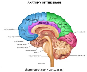
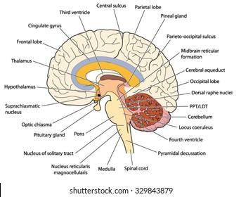
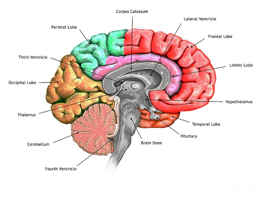
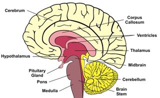
Brain cross section diagram
Brain Cross Section Diagram. Mobile and tablet users you can download e anatomy on appstore or googleplay. The brain s sensory switchboard located on top of the brainstem. Mri of the brain. All the functions are carried out without a single glitch and before you even bat an eyelid.
 Cross Section Brain Images Stock Photos Vectors Shutterstock From shutterstock.com
Of all the human body systems the nervous system is the most complicated system in the body. Mobile and tablet users you can download e anatomy on appstore or googleplay. We obtained 24 axial slices of the normal brain. Anatomical structures and specific areas are visible as interactive labeled images. It directs messages to the sensory receiving areas in the cortex and transmits replies to the cerebellum and medulla midbrain a small part of the brain above the pons that integrates sensory information and relays it upward. All the functions are carried out without a single glitch and before you even bat an eyelid.
Thousands of new high quality pictures added every day.
Mri of the brain. Sectional anatomy of the structures of the brain as viewed with ct mri and pet fusion imaging. This mri brain cross sectional anatomy tool is absolutely free to use. Mri of the brain. The human brain is an astonishing organ that takes care of each function and action of the body. The following are the different regions of the human brain and their functions.
 Source: shutterstock.com
We obtained 24 axial slices of the normal brain. Jul 3 2017 brain cross section diagram cross section of the human brain royalty free cliparts vectors photo brain cross section diagram cross section of the human brain royalty free cliparts vectors image brain cross section diagram cross section of the human brain royalty free cliparts vectors gallery. Mri of the brain. Anatomical structures and specific areas are visible as interactive labeled images. Of all the human body systems the nervous system is the most complicated system in the body.
 Source: fineartamerica.com
We obtained 24 axial slices of the normal brain. Mri of the brain. Spin echo t1 t2 and flair t2 gradient echo diffusion and t1 after gadolinium injection. It directs messages to the sensory receiving areas in the cortex and transmits replies to the cerebellum and medulla midbrain a small part of the brain above the pons that integrates sensory information and relays it upward. Thousands of new high quality pictures added every day.
 Source: flickr.com
Of all the human body systems the nervous system is the most complicated system in the body. The brain s sensory switchboard located on top of the brainstem. Jul 3 2017 brain cross section diagram cross section of the human brain royalty free cliparts vectors photo brain cross section diagram cross section of the human brain royalty free cliparts vectors image brain cross section diagram cross section of the human brain royalty free cliparts vectors gallery. If these are the questions swirling in your brain then this article detailing the diagram of the brain and its functions will definitely whet your appetite regarding brain functions and parts. Spin echo t1 t2 and flair t2 gradient echo diffusion and t1 after gadolinium injection.
 Source: livescience.com
Brain cross section diagram picture of brain cross section diagram. Webmd s brain anatomy page provides a detailed diagram and definition of the brain including its function parts and conditions that affect it. Anatomical structures and specific areas are visible as interactive labeled images. Spin echo t1 t2 and flair t2 gradient echo diffusion and t1 after gadolinium injection. This mri brain cross sectional anatomy tool is absolutely free to use.
 Source: sen842cova.blogspot.com
We obtained 24 axial slices of the normal brain. We obtained 24 axial slices of the normal brain. An mri was performed on a healthy subject with several acquisitions with different weightings. Anatomical structures and specific areas are visible as interactive labeled images. Labeled diagrams of the human brain.
 Source: pinterest.com
An mri was performed on a healthy subject with several acquisitions with different weightings. All the functions are carried out without a single glitch and before you even bat an eyelid. The human brain is an astonishing organ that takes care of each function and action of the body. Anatomical structures and specific areas are visible as interactive labeled images. Brain cross section diagram picture of brain cross section diagram.
 Source: shutterstock.com
The human brain is an astonishing organ that takes care of each function and action of the body. If these are the questions swirling in your brain then this article detailing the diagram of the brain and its functions will definitely whet your appetite regarding brain functions and parts. It directs messages to the sensory receiving areas in the cortex and transmits replies to the cerebellum and medulla midbrain a small part of the brain above the pons that integrates sensory information and relays it upward. The human brain is an astonishing organ that takes care of each function and action of the body. This mri brain cross sectional anatomy tool is absolutely free to use.
 Source: pinterest.com
Webmd s brain anatomy page provides a detailed diagram and definition of the brain including its function parts and conditions that affect it. If these are the questions swirling in your brain then this article detailing the diagram of the brain and its functions will definitely whet your appetite regarding brain functions and parts. An mri was performed on a healthy subject with several acquisitions with different weightings. This mri brain cross sectional anatomy tool is absolutely free to use. Find cross section of brain stock images in hd and millions of other royalty free stock photos illustrations and vectors in the shutterstock collection.
 Source: pinterest.com
An mri was performed on a healthy subject with several acquisitions with different weightings. Of all the human body systems the nervous system is the most complicated system in the body. Sectional anatomy of the structures of the brain as viewed with ct mri and pet fusion imaging. The brain s sensory switchboard located on top of the brainstem. Brain cross section diagram picture of brain cross section diagram.
 Source: quizlet.com
It directs messages to the sensory receiving areas in the cortex and transmits replies to the cerebellum and medulla midbrain a small part of the brain above the pons that integrates sensory information and relays it upward. We obtained 24 axial slices of the normal brain. An mri was performed on a healthy subject with several acquisitions with different weightings. The following are the different regions of the human brain and their functions. Brain cross section diagram picture of brain cross section diagram.
 Source: mpg.printstoreonline.com
Anatomical structures and specific areas are visible as interactive labeled images. Mri of the brain. Anatomical structures and specific areas are visible as interactive labeled images. The human brain is an astonishing organ that takes care of each function and action of the body. Sectional anatomy of the structures of the brain as viewed with ct mri and pet fusion imaging.
 Source: researchgate.net
Mobile and tablet users you can download e anatomy on appstore or googleplay. This mri brain cross sectional anatomy tool is absolutely free to use. The human brain is an astonishing organ that takes care of each function and action of the body. All the functions are carried out without a single glitch and before you even bat an eyelid. If these are the questions swirling in your brain then this article detailing the diagram of the brain and its functions will definitely whet your appetite regarding brain functions and parts.
 Source: quizlet.com
Spin echo t1 t2 and flair t2 gradient echo diffusion and t1 after gadolinium injection. Sectional anatomy of the structures of the brain as viewed with ct mri and pet fusion imaging. The human brain is an astonishing organ that takes care of each function and action of the body. Thousands of new high quality pictures added every day. Mobile and tablet users you can download e anatomy on appstore or googleplay.
 Source: radiopaedia.org
All the functions are carried out without a single glitch and before you even bat an eyelid. This mri brain cross sectional anatomy tool is absolutely free to use. The human brain is an astonishing organ that takes care of each function and action of the body. Jul 3 2017 brain cross section diagram cross section of the human brain royalty free cliparts vectors photo brain cross section diagram cross section of the human brain royalty free cliparts vectors image brain cross section diagram cross section of the human brain royalty free cliparts vectors gallery. Anatomical structures and specific areas are visible as interactive labeled images.
 Source: headinjury.com
If these are the questions swirling in your brain then this article detailing the diagram of the brain and its functions will definitely whet your appetite regarding brain functions and parts. Thousands of new high quality pictures added every day. It directs messages to the sensory receiving areas in the cortex and transmits replies to the cerebellum and medulla midbrain a small part of the brain above the pons that integrates sensory information and relays it upward. An mri was performed on a healthy subject with several acquisitions with different weightings. This mri brain cross sectional anatomy tool is absolutely free to use.
If you find this site adventageous, please support us by sharing this posts to your preference social media accounts like Facebook, Instagram and so on or you can also bookmark this blog page with the title brain cross section diagram by using Ctrl + D for devices a laptop with a Windows operating system or Command + D for laptops with an Apple operating system. If you use a smartphone, you can also use the drawer menu of the browser you are using. Whether it’s a Windows, Mac, iOS or Android operating system, you will still be able to bookmark this website.


