Cow eye dissection answers
Cow Eye Dissection Answers. Lab virtual eye dissection go to. Clic k dissection 101. There are three layers from the exterior. Choose from 500 different sets of cow eye dissection flashcards on quizlet.
 Pin By Angela Geiger Hutchison On Human Anatomy Drawing Human Eye Diagram Eye Anatomy Diagram Eye Anatomy From pinterest.com
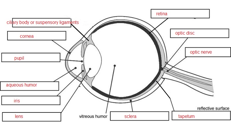
Lab virtual eye dissection go to. On the inside of the back half of the eyeball you can see some blood vessels that are part of a thin fleshy film. Obtain a cow eye place it in your dissecting pan rinse the eye with water. Step 1 the cow s eye 1. Tapetum mid layer of the eye and contains reflective pigments that helps the eye see better at night. Why does the exploratorium dissect cow eyes.
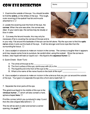
In the photo on the left is a picture of the intact eye and lying next to it is the excess superficial fatty tissue.
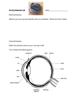
Krystina clark cow eye dissection observations exterior anatomy of the eye insert your photograph of the exterior of the intact eye here. Cow s eye dissection page 6 now take a look at the rest of the eye. Cow eye dissecting pan dissecting kit safety glasses lab apron and gloves. Click watch online answer the questions as you watch each video. L ens clear flexible structure that focuses the light on the retina. Keep cutting close to the sclera separating the membrane that attaches the muscle to it.
 Source: teacherspayteachers.com
Place the cow eye on a dissecting tray. What is one major difference between a cow s eye and a human s eye. These are the extrinsic muscles that allow a cow to move its eye up and down and from side to side. Cow eye use the point of a scissors or a scalpel to make an incision through the layers of the eye capsule similar to figure 1. In the photo on the left is a picture of the intact eye and lying next to it is the excess superficial fatty tissue.
 Source: exploratorium.edu
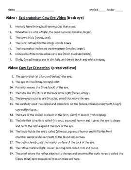
Click watch online answer the questions as you watch each video. Rotate the eye until the larger bulge or tear gland is on the top of the eye. Learn cow eye dissection with free interactive flashcards. Clic k dissection 101. What is one major difference between a cow s eye and a human s eye.
 Source: pinterest.com
Choose from 500 different sets of cow eye dissection flashcards on quizlet. Lab virtual eye dissection go to. Tapetum mid layer of the eye and contains reflective pigments that helps the eye see better at night. Clic k dissection 101. There are three layers from the exterior.
 Source: coursehero.com
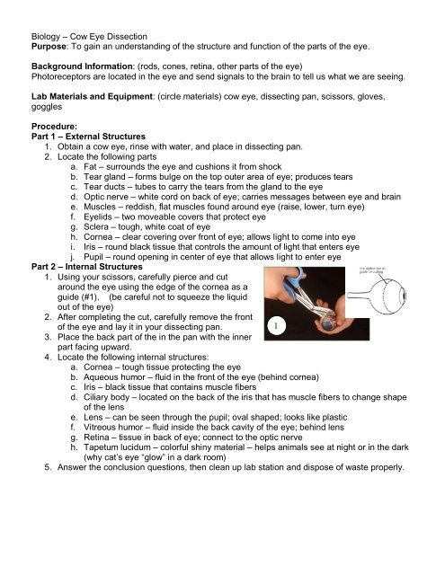
Cow eye use the point of a scissors or a scalpel to make an incision through the layers of the eye capsule similar to figure 1. If the vitreous humor is still in the eyeball empty it out. Clic k dissection 101. Cow eye dissecting pan dissecting kit safety glasses lab apron and gloves. The eye most likely has a thick covering of fat and muscle tissue.
 Source: studyres.com
Choose from 500 different sets of cow eye dissection flashcards on quizlet. Rotate the eye until the larger bulge or tear gland is on the top of the eye. Cow s eye dissection page 6 now take a look at the rest of the eye. Keep cutting close to the sclera separating the membrane that attaches the muscle to it. Step 1 the cow s eye 1.
Source:
Keep cutting close to the sclera separating the membrane that attaches the muscle to it. Why does the exploratorium dissect cow eyes. As you get closer to the actual eyeball you may notice muscles that are attached directly to the sclera and along the optic nerve. Blind spo t place where optic nerve leaves the retina. The eye most likely has a thick covering of fat and muscle tissue.
 Source: studylib.net
Tapetum mid layer of the eye and contains reflective pigments that helps the eye see better at night. Learn cow eye dissection with free interactive flashcards. What is one major difference between a cow s eye and a human s eye. L ens clear flexible structure that focuses the light on the retina. Step 1 the cow s eye 1.
 Source: studylib.net
What is one major difference between a cow s eye and a human s eye. Fovea centralis the point in the back of the eye with the greatest density of cone cells. That film is the retina. Lab virtual eye dissection go to. The eye most likely has a thick covering of fat and muscle tissue.
 Source: biologyjunction.com
Before you cut the eye open the vitreous humor. Learn cow eye dissection with free interactive flashcards. On the inside of the back half of the eyeball you can see some blood vessels that are part of a thin fleshy film. In the photo on the left is a picture of the intact eye and lying next to it is the excess superficial fatty tissue. Step 1 the cow s eye 1.
 Source: yumpu.com
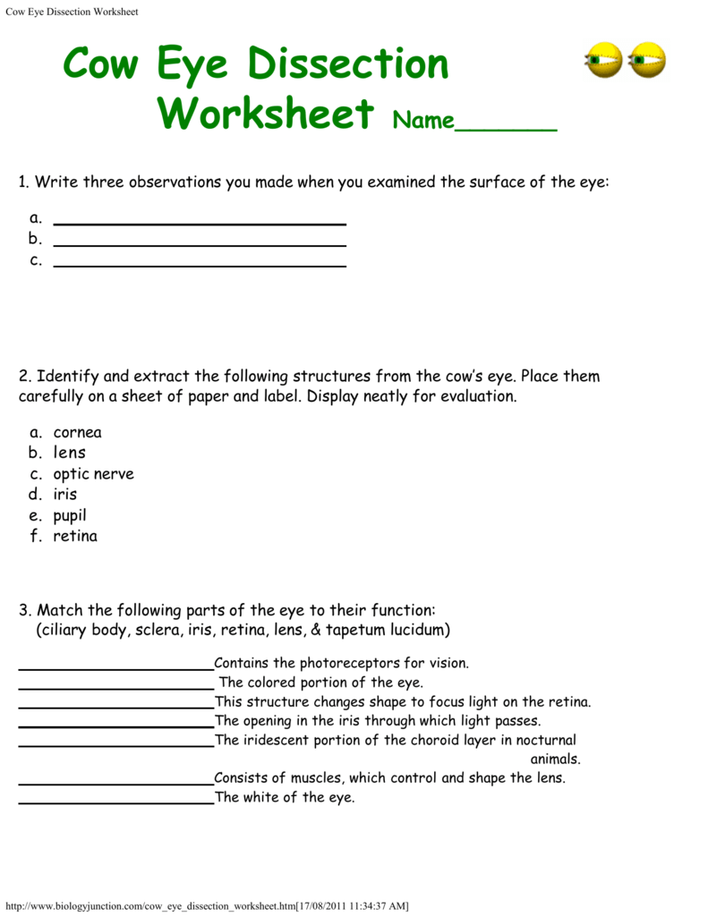
Include labels that identify the optic nerve the sclera and the cornea and a caption describing this view of the eye. Include labels that identify the optic nerve the sclera and the cornea and a caption describing this view of the eye. Choose from 500 different sets of cow eye dissection flashcards on quizlet. L ens clear flexible structure that focuses the light on the retina. Why does the exploratorium dissect cow eyes.
 Source: coursehero.com
Cow s eye dissection page 6 now take a look at the rest of the eye. Click watch online answer the questions as you watch each video. Rotate the eye until the larger bulge or tear gland is on the top of the eye. Why does the exploratorium dissect cow eyes. Step 1 the cow s eye 1.
 Source: biologycorner.com
There are three layers from the exterior. That film is the retina. Cow s eye dissection page 6 now take a look at the rest of the eye. Tapetum mid layer of the eye and contains reflective pigments that helps the eye see better at night. Lab virtual eye dissection go to.
 Source: biologycorner.com
Cow eye use the point of a scissors or a scalpel to make an incision through the layers of the eye capsule similar to figure 1. Learn cow eye dissection with free interactive flashcards. Carefully cut away the fat and the muscle. Cow eye dissecting pan dissecting kit safety glasses lab apron and gloves. These are the extrinsic muscles that allow a cow to move its eye up and down and from side to side.
 Source: promotiontablecovers.blogspot.com
Learn cow eye dissection with free interactive flashcards. Tapetum mid layer of the eye and contains reflective pigments that helps the eye see better at night. Choose from 500 different sets of cow eye dissection flashcards on quizlet. Fovea centralis the point in the back of the eye with the greatest density of cone cells. What is one major difference between a cow s eye and a human s eye.
 Source: studylib.net
Cow s eye dissection page 6 now take a look at the rest of the eye. In the photo on the left is a picture of the intact eye and lying next to it is the excess superficial fatty tissue. Fovea centralis the point in the back of the eye with the greatest density of cone cells. On the inside of the back half of the eyeball you can see some blood vessels that are part of a thin fleshy film. Sclera whitish grey continuous with the transparent cornea choroid thin dark black layer and the retina thin greyish pink layer use a scissors to.
If you find this site convienient, please support us by sharing this posts to your preference social media accounts like Facebook, Instagram and so on or you can also bookmark this blog page with the title cow eye dissection answers by using Ctrl + D for devices a laptop with a Windows operating system or Command + D for laptops with an Apple operating system. If you use a smartphone, you can also use the drawer menu of the browser you are using. Whether it’s a Windows, Mac, iOS or Android operating system, you will still be able to bookmark this website.


