Frog dissection organs
Frog Dissection Organs. Shows how the frog is cut to reveal the structures of the body cavity. You ll also see the glottis which is the opening to the lungs. Take care to cut only the skin. Video examines each of the main organs of the digestive system and then parts of the.
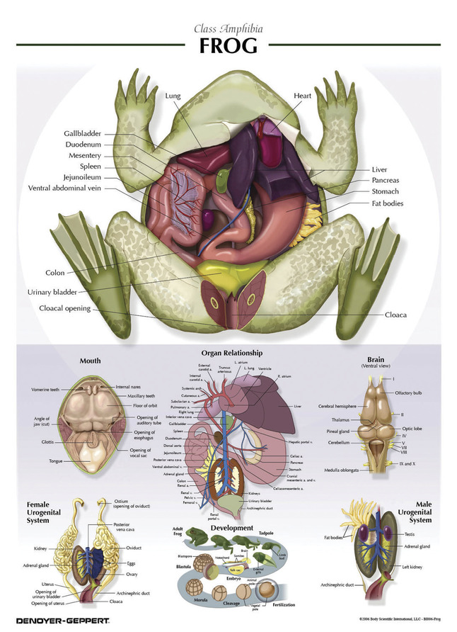 Denoyer Geppert Frog Anatomy Chart Laminated 21 X 29 Inches From schoolspecialty.com
Draw the frog s front foot and the back foot on the diagram. Place your frog on its back and pin it to the dissecting tray. The kidneys are organs that excrete urine. Front leg hind leg dorsal surface ventral surface mouth upper eyelid lower eyelid. This organ is the heart. Place the frog in the dissecting pan ventral side up.
Frog organs and functions all about our class dissection.
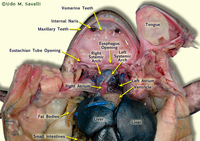
Carefully cut away the pericardium the thin membrane surrounding the heart. Carefully cut away the pericardium the thin membrane surrounding the heart. The kidneys are organs that excrete urine. Internal organs 3 dimensional. Mouthinside the frog s mouth you will see the esophagus a tube where food passes from the mouth to the stomach. This organ is the heart.
 Source: ez002.k12.sd.us
The simulation includes the following sections. Its brown in color and consists of lobes. Remember there is a difference in a frog s front and back feet. Use this printable frog dissection diagram with labeled parts pdf as a guide for locating them. Internal organs 3 dimensional.
 Source: pinterest.com
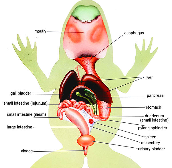
You will write in the length of these legs during the lab. Use this printable frog dissection diagram with labeled parts pdf as a guide for locating them. This organ is the liver. Front leg hind leg dorsal surface ventral surface mouth upper eyelid lower eyelid. The urinary system consists of the frog s kidneys ureters bladder and cloaca.
 Source: amazon.com
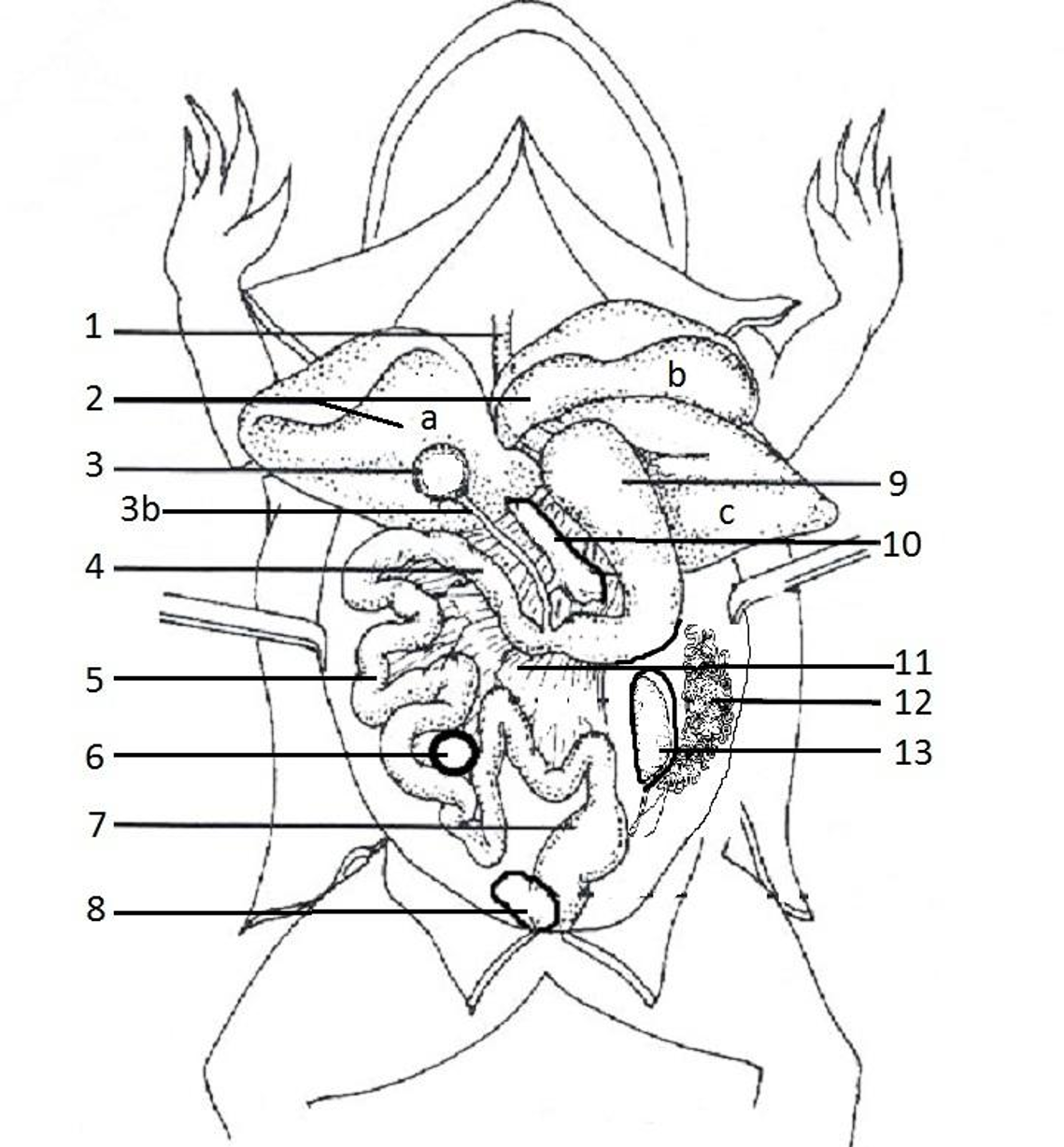
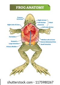
Shows how the frog is cut to reveal the structures of the body cavity. Video examines each of the main organs of the digestive system and then parts of the. Shows how the frog is cut to reveal the structures of the body cavity. The liver is the largest structure of the body cavity. Use this printable frog dissection diagram with labeled parts pdf as a guide for locating them.
 Source: pinterest.com
Place the frog in the dissecting pan ventral side up. The simulation includes the following sections. The kidneys are organs that excrete urine. Shows how the frog is cut to reveal the structures of the body cavity. Frog organs and functions all about our class dissection.
 Source: pinterest.com
Mouthinside the frog s mouth you will see the esophagus a tube where food passes from the mouth to the stomach. The frog dissection is a simulation app of wet lab dissection procedures. Make a small cut through the lifted skin with the scalpel. Shows how the frog is cut to reveal the structures of the body cavity. Cut along the midline of the body to the forelimbs.
 Source: schoolspecialty.com
Place the frog in the dissecting pan ventral side up. Carefully cut away the pericardium the thin membrane surrounding the heart. The simulation includes the following sections. Lift the frog s skin with forceps between the rear legs. The kidneys are organs that excrete urine.
 Source: biologycorner.com
Carefully cut away the pericardium the thin membrane surrounding the heart. Use scissors to lift the abdominal muscles away from the body cavity. The liver is the largest structure of the body cavity. Internal organs 3 dimensional. Remember there is a difference in a frog s front and back feet.
 Source: pinterest.com
Take care to cut only the skin. Use scissors to lift the abdominal muscles away from the body cavity. You ll also see the glottis which is the opening to the lungs. This is a starting place for the scissors. Food is moved down the esophagus by a process called peristalsis which is coordinated contraction of muscles in the esophagus.
 Source: study.com
Shows how the frog is cut to reveal the structures of the body cavity. Mouthinside the frog s mouth you will see the esophagus a tube where food passes from the mouth to the stomach. Carefully cut away the pericardium the thin membrane surrounding the heart. You ll also see the glottis which is the opening to the lungs. The liver s function is to make a digestive juice named bile and its needed for the digestion of fats.
 Source: pinterest.com
Draw the frog s front foot and the back foot on the diagram. Use scissors to lift the abdominal muscles away from the body cavity. You will write in the length of these legs during the lab. Place the frog in the dissecting pan ventral side up. Mouthinside the frog s mouth you will see the esophagus a tube where food passes from the mouth to the stomach.
 Source: savalli.us
The kidneys are organs that excrete urine. The liver is the largest structure of the body cavity. Video examines each of the main organs of the digestive system and then parts of the. This organ is the heart. You will write in the length of these legs during the lab.
 Source: shutterstock.com
Draw the frog s front foot and the back foot on the diagram. Connected to each kidney is a ureter a tube through which urine passes into the urinary bladder a sac that stores urine until it passes out of the body through the cloaca. This organ is the heart. Video examines each of the main organs of the digestive system and then parts of the. You will write in the length of these legs during the lab.
 Source: pinterest.com
Shows how the frog is cut to reveal the structures of the body cavity. Unlike a mammal heart it only has three chambers two atria at the top and one ventricle below. Cut along the midline of the body to the forelimbs. The liver is the largest structure of the body cavity. Frog organs and functions all about our class dissection.
 Source: quizlet.com
Frog dissection pdf australian curriculum alignment multicellular organisms have a hierarchical structural organisation of cells tissues organs and systems acsbl054. This is a starting place for the scissors. The urinary system consists of the frog s kidneys ureters bladder and cloaca. The frog dissection is a simulation app of wet lab dissection procedures. Internal organs 3 dimensional.
 Source: biologycorner.com
Connected to each kidney is a ureter a tube through which urine passes into the urinary bladder a sac that stores urine until it passes out of the body through the cloaca. The liver is the largest structure of the body cavity. Connected to each kidney is a ureter a tube through which urine passes into the urinary bladder a sac that stores urine until it passes out of the body through the cloaca. Use scissors to lift the abdominal muscles away from the body cavity. The frog dissection is a simulation app of wet lab dissection procedures.
If you find this site adventageous, please support us by sharing this posts to your preference social media accounts like Facebook, Instagram and so on or you can also save this blog page with the title frog dissection organs by using Ctrl + D for devices a laptop with a Windows operating system or Command + D for laptops with an Apple operating system. If you use a smartphone, you can also use the drawer menu of the browser you are using. Whether it’s a Windows, Mac, iOS or Android operating system, you will still be able to bookmark this website.

