Heart anatomy diagram label
Heart Anatomy Diagram Label. Drag and drop the text labels onto the boxes next to the heart diagram. A heart diagram is a popular design used by different people for various uses. The human heart is situated under the ribcage. In this interactive you can label parts of the human heart.
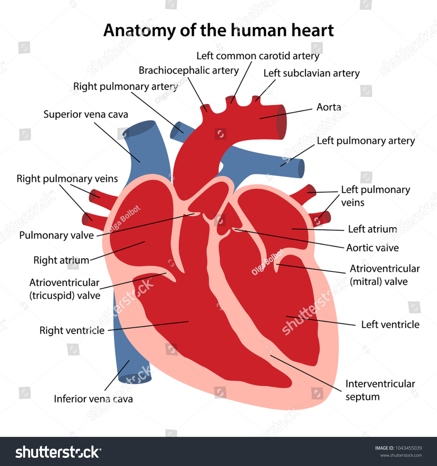 Anatomy Human Heart Cross Sectional Diagram Stock Vector Royalty Free 1043455039 From shutterstock.com
Oxygen poor blood enters the right atrium of the heart via veins called the inferior vena cava and the superior vena cava. Heart diagram is the outlook figure of heart. In this interactive you can label parts of the human heart. The heart sits within a fluid filled cavity called the pericardial cavity. Webmd s heart anatomy page provides a detailed image of the heart and provides information on heart conditions tests and treatments. Drag and drop the text labels onto the boxes next to the heart diagram.
Figure 1 shows the position of the heart within the thoracic cavity.
The areas of the heart with less oxygen are labeled with a b. In this interactive you can label parts of the human heart. Location of the heart. The blood is then pumped into the right ventricle and then through the pulmonary artery to the lungs where the blood is enriched with oxygen. The walls and lining of the pericardial cavity are a special membrane known as the pericardium. Heart diagram is the outlook figure of heart.
 Source: teachervision.com
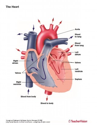
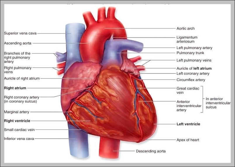
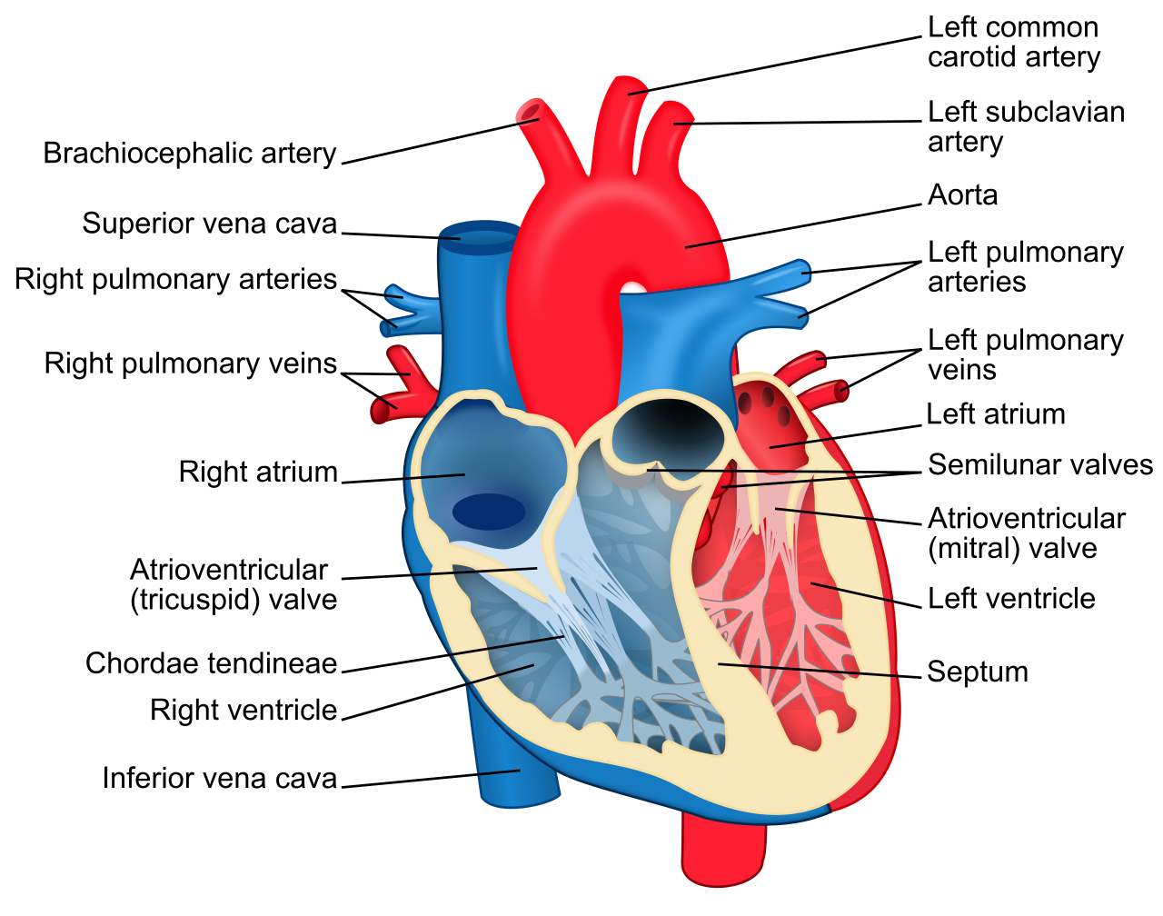
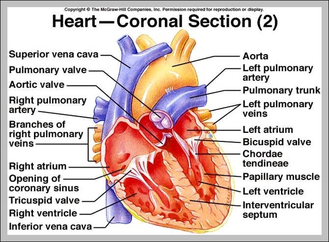
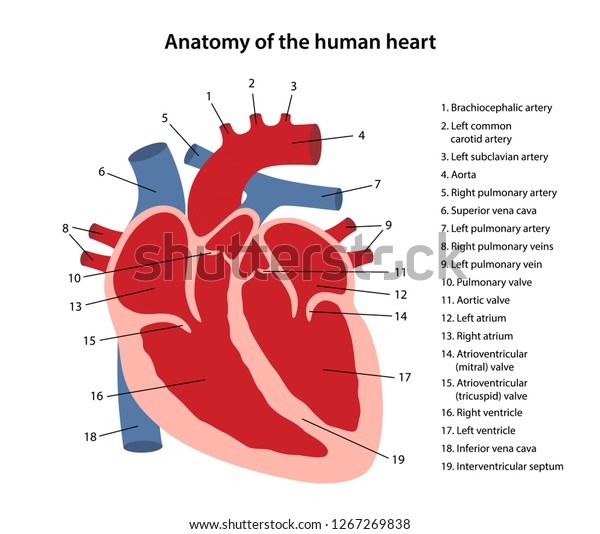
The septum separates the ventricles from each other and can be seen in the labeled heart diagram. A heart diagram labeled will provide plenty of information about the structure of your heart including the wall of your heart. If you want to redo an answer click on the box and the answer will go back to the top so you can move it to another box. Label heart interior anatomy diagram human anatomy the heart is a fist sized muscular organ that pumps blood through the body. The diagrams are provided in colors to show different parts of important heart component such as the aorta pulmonary artery pulmonary vein left atrium right atrium left ventricle right ventricle inferior vena cava and superior vena cava among others.
 Source: pinterest.com
The diagrams are provided in colors to show different parts of important heart component such as the aorta pulmonary artery pulmonary vein left atrium right atrium left ventricle right ventricle inferior vena cava and superior vena cava among others. The heart has four different chambers. Anatomy of the heart. The areas of the heart with less oxygen are labeled with a b. Figure 1 shows the position of the heart within the thoracic cavity.
 Source: tartrerepub.blogspot.com
The walls and lining of the pericardial cavity are a special membrane known as the pericardium. The human heart is located within the thoracic cavity medially between the lungs in the space known as the mediastinum. The left and right ventricles and the left and right atriums the chambers of the heart and the valves that regulate blood flow to them are considered the plumbing of the heart. A heart diagram labeled will provide plenty of information about the structure of your heart including the wall of your heart. Drag and drop the text labels onto the boxes next to the heart diagram.
 Source: kenhub.com
The walls and lining of the pericardial cavity are a special membrane known as the pericardium. Observe the following labeled heart diagrams to help you comprehend the different parts of the human heart. The blood is then pumped into the right ventricle and then through the pulmonary artery to the lungs where the blood is enriched with oxygen. Figure 1 shows the position of the heart within the thoracic cavity. The heart has four different chambers.
 Source: pinterest.com
Heart diagram is the outlook figure of heart. Its major function is pumping blood continually muscular wall beat or contraction and blood pumping to all the parts of the body. Anatomy of the heart. If you want to redo an answer click on the box and the answer will go back to the top so you can move it to another box. The human heart usually weighs somewhere between 10 to 12 ounces in men and between 8 to 10 ounces in women and in terms of size is roughly the size of the fist.
 Source: en.wikipedia.org
Anatomy of the heart. In the human heart diagram two atria and ventricles are separated from each other by a muscle wall called septum. In order to understand how that happens it is necessary to understand the anatomy and physiology of the heart. Heart diagram is the outlook figure of heart. The left and right ventricles and the left and right atriums the chambers of the heart and the valves that regulate blood flow to them are considered the plumbing of the heart.
 Source: shutterstock.com
The human heart is situated under the ribcage. The human heart is situated under the ribcage. Anatomy of the heart pericardium. The septum separates the ventricles from each other and can be seen in the labeled heart diagram. A heart diagram is a popular design used by different people for various uses.
 Source: anatomysystem.com
The areas of the heart with less oxygen are labeled with a b. Drag and drop the text labels onto the boxes next to the heart diagram. The blood is then pumped into the right ventricle and then through the pulmonary artery to the lungs where the blood is enriched with oxygen. In the human heart diagram two atria and ventricles are separated from each other by a muscle wall called septum. Anatomy of the heart.
 Source: tartrerepub.blogspot.com
The left and right ventricles and the left and right atriums the chambers of the heart and the valves that regulate blood flow to them are considered the plumbing of the heart. The walls and lining of the pericardial cavity are a special membrane known as the pericardium. Anatomy of the heart. Observe the following labeled heart diagrams to help you comprehend the different parts of the human heart. Webmd s heart anatomy page provides a detailed image of the heart and provides information on heart conditions tests and treatments.

The diagrams are provided in colors to show different parts of important heart component such as the aorta pulmonary artery pulmonary vein left atrium right atrium left ventricle right ventricle inferior vena cava and superior vena cava among others. Heres more about these three layers. Label heart interior anatomy diagram human anatomy the heart is a fist sized muscular organ that pumps blood through the body. Anatomy of the heart pericardium. Because the heart points to the left about 2 3 of the heart s mass is found on the left side of the body and the other 1 3 is on the right.
 Source: pinterest.com
Heart diagram is the outlook figure of heart. Figure 1 shows the position of the heart within the thoracic cavity. The human heart is situated under the ribcage. Heart diagram is the outlook figure of heart. It can be used by a teacher or student for academic purpose by a friend or relative for mutually sending and exchanging cards or for baby toys or printing on dresses etc.
 Source: pinterest.com
Anatomy of the heart pericardium. The human heart is situated under the ribcage. Label heart interior anatomy diagram human anatomy the heart is a fist sized muscular organ that pumps blood through the body. Webmd s heart anatomy page provides a detailed image of the heart and provides information on heart conditions tests and treatments. The blood is then pumped into the right ventricle and then through the pulmonary artery to the lungs where the blood is enriched with oxygen.
 Source: shutterstock.com
In this interactive you can label parts of the human heart. 0 0000 a shoutout is a way of letting people know of a game you want them to play. It can be used by a teacher or student for academic purpose by a friend or relative for mutually sending and exchanging cards or for baby toys or printing on dresses etc. The heart sits within a fluid filled cavity called the pericardial cavity. Its major function is pumping blood continually muscular wall beat or contraction and blood pumping to all the parts of the body.
 Source: researchgate.net
The heart has four different chambers. The heart has four different chambers. The areas of the heart with less oxygen are labeled with a b. Heres more about these three layers. 0 0000 a shoutout is a way of letting people know of a game you want them to play.
 Source: pinterest.com
Its major function is pumping blood continually muscular wall beat or contraction and blood pumping to all the parts of the body. Oxygen poor blood enters the right atrium of the heart via veins called the inferior vena cava and the superior vena cava. A heart diagram labeled will provide plenty of information about the structure of your heart including the wall of your heart. Location of the heart. If you want to redo an answer click on the box and the answer will go back to the top so you can move it to another box.
If you find this site value, please support us by sharing this posts to your favorite social media accounts like Facebook, Instagram and so on or you can also save this blog page with the title heart anatomy diagram label by using Ctrl + D for devices a laptop with a Windows operating system or Command + D for laptops with an Apple operating system. If you use a smartphone, you can also use the drawer menu of the browser you are using. Whether it’s a Windows, Mac, iOS or Android operating system, you will still be able to bookmark this website.


