Onion root tip mitosis slides labeled
Onion Root Tip Mitosis Slides Labeled. Then another onion root tip was prepared and area z was located. First with a prepared slide area x and y were located and each counted and recorded of what stages were observed. Microtubules align chromosomes along metaphase plate. Mitosis in onion root tip.
 Anatomy A215 Virtual Microscopy From medsci.indiana.edu
Observed in each part of mitosis for different areas of an onion root. Start studying onion root tip mitosis slide. Prepared microscope slide of a whitefish embryo. Mitosis in onion root tip. Focus in on low power and then switch to medium or high power. Based on the kind of cells and species of organism the time taken for mitosis may vary.
4 whitefish blastula mitosis this from the first stages of a fish embryo.
Preparing the root tip squash. First with a prepared slide area x and y were located and each counted and recorded of what stages were observed. Then another onion root tip was prepared and area z was located. 4 whitefish blastula mitosis this from the first stages of a fish embryo. Prepared microscope slide of an onion root tip. The meristamatic cells located in the root tips provide the most suitable material for the study of mitosis.
 Source: chegg.com
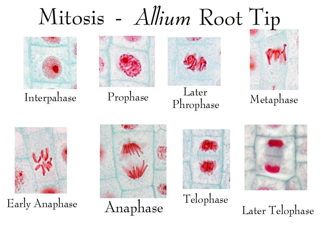
Onion root tip cell mitosis. Transfer a root to the center of a clean microscope slide and add a drop of water. Preparing the root tip squash. Below find micrographs of the four stages of mitosis. Focus as desired to obtain a distinct and clear image.
 Source: medsci.indiana.edu
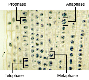
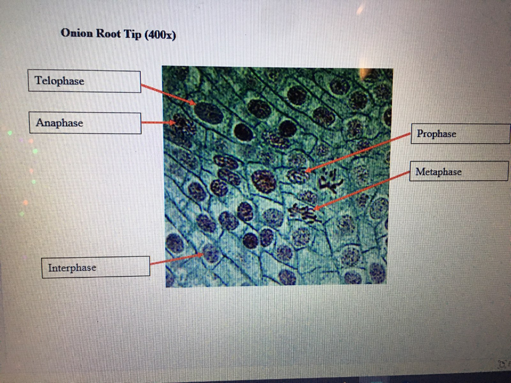
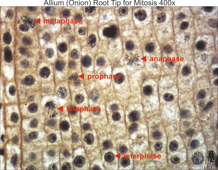
With the data of all three areas the mitotic stages were able to be easily analyzed. Prepared microscope slide of an onion root tip. When observing the onion root tip cells for the stage of prophase the cells took on a brick like structure and within the cells small dots the nuclei can be seen. Nuclear membrane breaks down chromatin condenses mitotic spindle forms and attaches to kinetochores. Mitosis in onion root tip.
 Source: bio.libretexts.org
The meristamatic cells located in the root tips provide the most suitable material for the study of mitosis. Prepared microscope slide of an onion root tip. Preparing the root tip squash. Using a razor blade cut off most of the unstained part of the root and discard it. The meristamatic cells located in the root tips provide the most suitable material for the study of mitosis.
 Source: pinterest.com
Place a slide containing a stained preparation of the allium onion root tip or whitefish blastula. Start studying onion root tip mitosis slide. Observed in each part of mitosis for different areas of an onion root. Using a razor blade cut off most of the unstained part of the root and discard it. Focus in on low power and then switch to medium or high power.
 Source: slideshare.net
Focus in on low power and then switch to medium or high power. Start studying onion root tip mitosis slide. 2 meiosis in ascaris meiosis ii chromosomes polar body meiosis in ascaris polar body chromosomes. Focus as desired to obtain a distinct and clear image. When observing the onion root tip cells for the stage of prophase the cells took on a brick like structure and within the cells small dots the nuclei can be seen.
 Source: search.library.wisc.edu
Microtubules align chromosomes along metaphase plate. Mitosis in onion root tip. 2 meiosis in ascaris meiosis ii chromosomes polar body meiosis in ascaris polar body chromosomes. The chromosome of monocotyledonous plants is large and more visible therefore onion root tips are used to study mitosis. Start studying onion root tip mitosis slide.
 Source: slideplayer.com
Below find micrographs of the four stages of mitosis. With the data of all three areas the mitotic stages were able to be easily analyzed. 4 whitefish blastula mitosis this from the first stages of a fish embryo. Locate the meristematic or growth zone which is just above the root cap at the very end of the tip or 4. Microtubules align chromosomes along metaphase plate.
 Source: in.pinterest.com
Prepared microscope slide of a whitefish embryo. Locate the meristematic or growth zone which is just above the root cap at the very end of the tip or 4. Place a slide containing a stained preparation of the allium onion root tip or whitefish blastula. Start studying onion root tip mitosis slide. Based on the kind of cells and species of organism the time taken for mitosis may vary.
 Source: biology.arizona.edu
Focus in on low power and then switch to medium or high power. Learn vocabulary terms and more with flashcards games and other study tools. Using a razor blade cut off most of the unstained part of the root and discard it. Based on the kind of cells and species of organism the time taken for mitosis may vary. Cover the root tip with a cover slip and then carefully push down on the cover slide with the.
 Source: pinterest.com
Prepared microscope slide of a whitefish embryo. 2 meiosis in ascaris meiosis ii chromosomes polar body meiosis in ascaris polar body chromosomes. Onion root tip cell mitosis. The chromosome of monocotyledonous plants is large and more visible therefore onion root tips are used to study mitosis. When observing the onion root tip cells for the stage of prophase the cells took on a brick like structure and within the cells small dots the nuclei can be seen.
 Source: laboratoriorojan.com.br
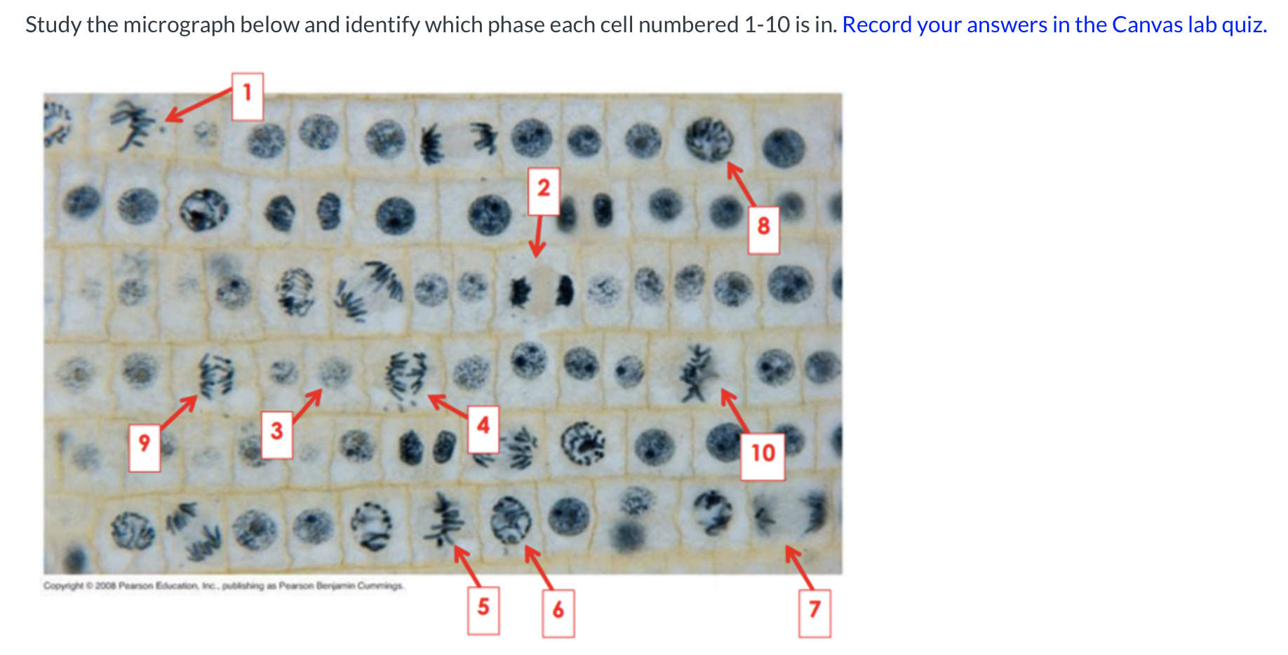
Transfer a root to the center of a clean microscope slide and add a drop of water. Focus in on low power and then switch to medium or high power. Transfer a root to the center of a clean microscope slide and add a drop of water. Microtubules align chromosomes along metaphase plate. Mitosis in onion root tip mitotic figures in onion root tip higher magnification than previous slide.
 Source: chegg.com
The meristamatic cells located in the root tips provide the most suitable material for the study of mitosis. Mitosis in onion root tip mitotic figures in onion root tip higher magnification than previous slide. Learn vocabulary terms and more with flashcards games and other study tools. Nuclear membrane breaks down chromatin condenses mitotic spindle forms and attaches to kinetochores. When observing the onion root tip cells for the stage of prophase the cells took on a brick like structure and within the cells small dots the nuclei can be seen.
 Source: pinterest.com
The chromosome of monocotyledonous plants is large and more visible therefore onion root tips are used to study mitosis. 4 whitefish blastula mitosis this from the first stages of a fish embryo. The chromosome of monocotyledonous plants is large and more visible therefore onion root tips are used to study mitosis. The meristamatic cells located in the root tips provide the most suitable material for the study of mitosis. When observing the onion root tip cells for the stage of prophase the cells took on a brick like structure and within the cells small dots the nuclei can be seen.
 Source: quizlet.com
The chromosome of monocotyledonous plants is large and more visible therefore onion root tips are used to study mitosis. 3 chromosomes grasshopper testis mature sperm cells grasshopper testis. Mitosis in onion root tip. Learn vocabulary terms and more with flashcards games and other study tools. 4 whitefish blastula mitosis this from the first stages of a fish embryo.
 Source: dissectionconnection.com.au
Onion root tip cell mitosis. The meristamatic cells located in the root tips provide the most suitable material for the study of mitosis. Based on the kind of cells and species of organism the time taken for mitosis may vary. When observing the onion root tip cells for the stage of prophase the cells took on a brick like structure and within the cells small dots the nuclei can be seen. Transfer a root to the center of a clean microscope slide and add a drop of water.
If you find this site good, please support us by sharing this posts to your favorite social media accounts like Facebook, Instagram and so on or you can also bookmark this blog page with the title onion root tip mitosis slides labeled by using Ctrl + D for devices a laptop with a Windows operating system or Command + D for laptops with an Apple operating system. If you use a smartphone, you can also use the drawer menu of the browser you are using. Whether it’s a Windows, Mac, iOS or Android operating system, you will still be able to bookmark this website.


