Sheep heart with labels
Sheep Heart With Labels. All the major vessels are represented many are labeled with colored pencils so that you can see exactly where each is located. Label the valves pdf internal anatomy. This page contains photos of the sheep heart dissection. Sheep heart dissection sheep have a four chambered heart just like humans.

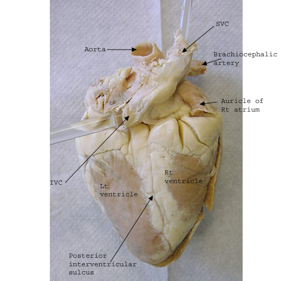
This image shows an external view of the anterior side of a preserved sheep heart. Label the valves pdf internal anatomy. The heart can be confusing because it is not perfectly symmetrical. Label the parts of a sheep heart. All the major vessels are represented many are labeled with colored pencils so that you can see exactly where each is located. Note the pointed apex of the heart and the wide superior end of the heart which is termed the base.
All the major vessels are represented many are labeled with colored pencils so that you can see exactly where each is located.
This page contains photos of the sheep heart dissection. The wall of the right ventricle will be significantly thinner. The visceral layer of the pericardium the epicardium is attached to the outer surface of the heart wall. Label the left side pdf see our other free dissection guides with photos and printable pdfs. As you view this set be mindful of how the heart was bisected look for clues example. The heart is suspended in a double walled fibroserous sac called the pericardium.
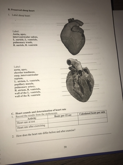
By studying the anatomy of a sheep s heart you can learn about how your own heart pumps blood through your body and keeps you alive. The visceral layer of the pericardium the epicardium is attached to the outer surface of the heart wall. Students often confuse the left and the right side of the heart. All the major vessels are represented many are labeled with colored pencils so that you can see exactly where each is located. This image shows an external view of the anterior side of a preserved sheep heart.

Label the right side pdf internal anatomy. The heart can be confusing because it is not perfectly symmetrical. Label the valves pdf internal anatomy. Use this as a dissection guide complete enough for a high school lab or just look at the labeled images to. Label the left side pdf see our other free dissection guides with photos and printable pdfs.
 Source: classroom.sdmesa.edu
This image shows an external view of the anterior side of a preserved sheep heart. Label the left side pdf see our other free dissection guides with photos and printable pdfs. Sheep heart dissection sheep have a four chambered heart just like humans. All the major vessels are represented many are labeled with colored pencils so that you can see exactly where each is located. Students often confuse the left and the right side of the heart.

This image shows an external view of the anterior side of a preserved sheep heart. Print out these diagrams and fill in the labels to test your knowledge of sheep heart anatomy. Students often confuse the left and the right side of the heart. Label the right side pdf internal anatomy. As you view this set be mindful of how the heart was bisected look for clues example.
 Source: pinterest.com
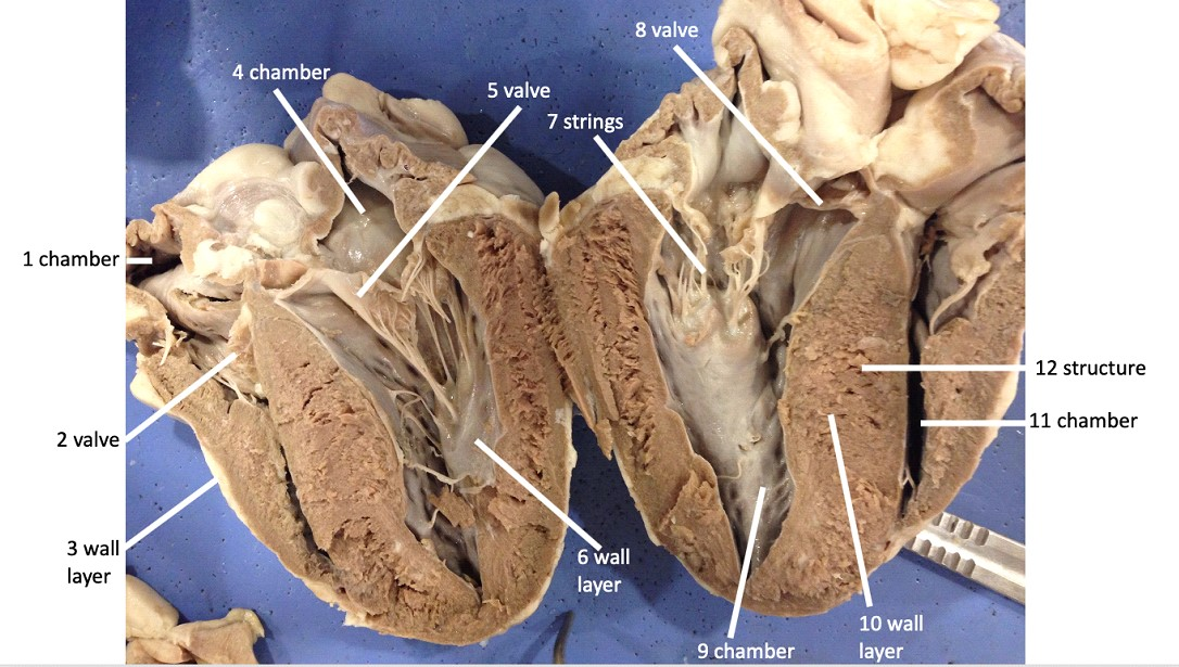
The incision should follow the line of the right side of the heart so that you can open just the right side and see the right atrium the right ventricle and the tricuspid valve between them. The large blood vessels i e the great vessels of the heart which carry blood to and from the heart are located at the base. The visceral layer of the pericardium the epicardium is attached to the outer surface of the heart wall. Students often confuse the left and the right side of the heart. Sheep heart dissection sheep have a four chambered heart just like humans.
Source:
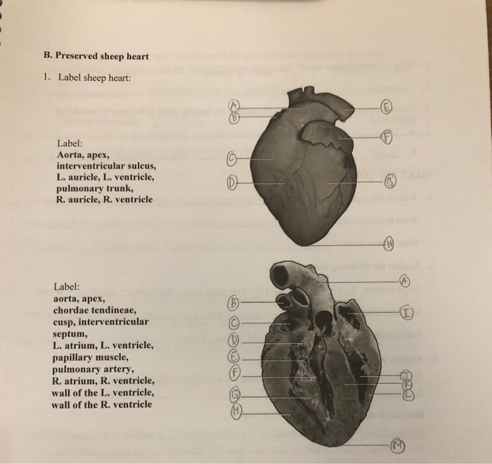
The large blood vessels i e the great vessels of the heart which carry blood to and from the heart are located at the base. Print out these diagrams and fill in the labels to test your knowledge of sheep heart anatomy. Use a scalpel to make an incision in the heart at the superior vena cava. Sheep heart dissection sheep have a four chambered heart just like humans. Label the parts of a sheep heart.
 Source: chegg.com
Students often confuse the left and the right side of the heart. The heart can be confusing because it is not perfectly symmetrical. Note the pointed apex of the heart and the wide superior end of the heart which is termed the base. By studying the anatomy of a sheep s heart you can learn about how your own heart pumps blood through your body and keeps you alive. Label the left side pdf see our other free dissection guides with photos and printable pdfs.
 Source: carolina.com
Print out these diagrams and fill in the labels to test your knowledge of sheep heart anatomy. The incision should follow the line of the right side of the heart so that you can open just the right side and see the right atrium the right ventricle and the tricuspid valve between them. The wall of the right ventricle will be significantly thinner. The heart is suspended in a double walled fibroserous sac called the pericardium. Made with explain everything.
 Source: classroom.sdmesa.edu
This image shows an external view of the anterior side of a preserved sheep heart. Label the right side pdf internal anatomy. Sheep heart dissection sheep have a four chambered heart just like humans. As you view this set be mindful of how the heart was bisected look for clues example. By studying the anatomy of a sheep s heart you can learn about how your own heart pumps blood through your body and keeps you alive.
 Source: chegg.com
This image shows an external view of the anterior side of a preserved sheep heart. The heart can be confusing because it is not perfectly symmetrical. Print out these diagrams and fill in the labels to test your knowledge of sheep heart anatomy. Label the right side pdf internal anatomy. Label the left side pdf see our other free dissection guides with photos and printable pdfs.
 Source: br.pinterest.com
This image shows an external view of the anterior side of a preserved sheep heart. The incision should follow the line of the right side of the heart so that you can open just the right side and see the right atrium the right ventricle and the tricuspid valve between them. Use this as a dissection guide complete enough for a high school lab or just look at the labeled images to. The heart can be confusing because it is not perfectly symmetrical. The heart is suspended in a double walled fibroserous sac called the pericardium.
 Source: quizlet.com
The majority of this sac is usually absent from the prepared sheep hearts but there may be parts of it still attached to the great vessels of the heart. Sheep heart dissection sheep have a four chambered heart just like humans. This page contains photos of the sheep heart dissection. Print out these diagrams and fill in the labels to test your knowledge of sheep heart anatomy. 1 identify the right and left sides of the heart look closely and on one side you will see a diagonal line of blood vessels that divide the heart this line is called the interventricular sulcus the half that includes all of the apex pointed end of the heart is the left side.
 Source: pinterest.com
Label the left side pdf see our other free dissection guides with photos and printable pdfs. Students often confuse the left and the right side of the heart. The majority of this sac is usually absent from the prepared sheep hearts but there may be parts of it still attached to the great vessels of the heart. Label the right side pdf internal anatomy. Note the pointed apex of the heart and the wide superior end of the heart which is termed the base.
 Source: quizlet.com
Label the parts of a sheep heart. The wall of the right ventricle will be significantly thinner. Sheep heart dissection sheep have a four chambered heart just like humans. Label the parts of a sheep heart. This image shows an external view of the anterior side of a preserved sheep heart.
 Source: carolina.com
Label the valves pdf internal anatomy. Use a scalpel to make an incision in the heart at the superior vena cava. All the major vessels are represented many are labeled with colored pencils so that you can see exactly where each is located. The visceral layer of the pericardium the epicardium is attached to the outer surface of the heart wall. This page contains photos of the sheep heart dissection.
If you find this site value, please support us by sharing this posts to your own social media accounts like Facebook, Instagram and so on or you can also save this blog page with the title sheep heart with labels by using Ctrl + D for devices a laptop with a Windows operating system or Command + D for laptops with an Apple operating system. If you use a smartphone, you can also use the drawer menu of the browser you are using. Whether it’s a Windows, Mac, iOS or Android operating system, you will still be able to bookmark this website.

