Sheep pluck dissection instructions
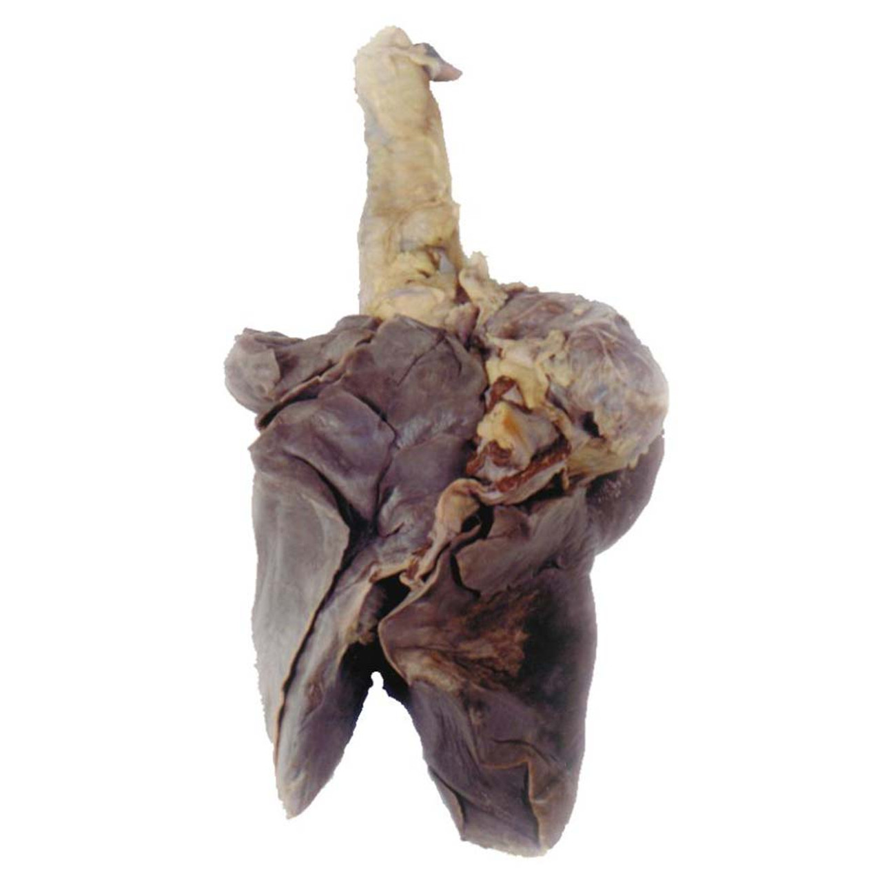
Sheep Pluck Dissection Instructions. This resource was funded by the gatsby charitable foundation and. Distribute the instruments to students. Go back to the top of the bronchiole use your scissors to cut down one bronchus following it through the lung. Aprons and goggles are also available for additional protection.
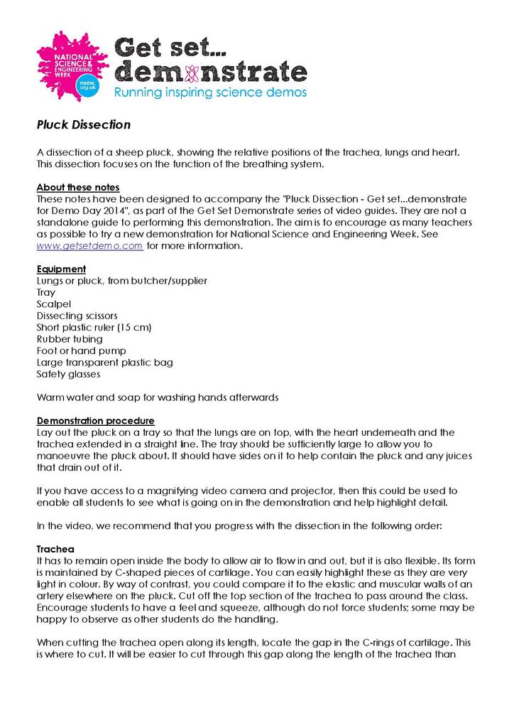 Pluck Dissection Stem From stem.org.uk
Distribute the instruments to students. Right atrium lungs part 1 heart dissection the right upper chamber of the heart. Place disinfectant in a container ready for instruments to be placed at the end of the dissection. Into the superior vena cava and make an incision down through the wall of the right atrium and ventricle as shown by the dotted line in the external heart picture. When handling the sheep s pluck make sure to wear disposable gloves. Go back to the top of the bronchiole use your scissors to cut down one bronchus following it through the lung.
Prepare disinfectant solution according to manufacturer s instructions.
It is the part of the heart that receives the deoxygenated blood from the whole body through the venae cavae. Take care to clearly illustrate and label the four lobes. As seen in the picture to the left. This should be a clear lump which feels soft on the outside and hard in the middle. When handling the sheep s pluck make sure to wear disposable gloves. Aprons and goggles are also available for additional protection.
 Source: coursehero.com
As seen in the picture to the left. Take care to clearly illustrate and label the four lobes. If any blood is associated with the lungs or pluck rinse them in cold running water. Cut a piece of the lung tissue out and you should be able to see the tubes branching out. Remember to draw your illustrations to scale.
 Source: stem.org.uk
Insert your dissecting scissors or scalpel. These are called semi lunar valves and prevent blood from flowing backwards from the arteries. Click for full size pdf. This resource was funded by the gatsby charitable foundation and. Distribute the instruments to students.
Source:
Into the superior vena cava and make an incision down through the wall of the right atrium and ventricle as shown by the dotted line in the external heart picture. Aprons and goggles are also available for additional protection. This should be a clear lump which feels soft on the outside and hard in the middle. Into the superior vena cava and make an incision down through the wall of the right atrium and ventricle as shown by the dotted line in the external heart picture. These are called semi lunar valves and prevent blood from flowing backwards from the arteries.
Source:
This video demonstrates the dissection of a sheep s heart in a laboratory setting. Shows the completed dissection of the sheep heart. Distribute the instruments to students. This should be a clear lump which feels soft on the outside and hard in the middle. The retina should be attached only at one point of the eye.
Source:
Ensure students have appropriate ppe. Aprons and goggles are also available for additional protection. As seen in the picture to the left. The retina should be attached only at one point of the eye. Insert your dissecting scissors or scalpel.
 Source: evolvingsciences.com
Operating procedure cont figure 1. Prepare disinfectant solution according to manufacturer s instructions. Draw a picture of the left lung of the sheep. Now examine the back half of the eye you should be able to see some thin blood vessels that are part of a thin fl eshy fi lm this fi lm is the retina. Go back to the top of the bronchiole use your scissors to cut down one bronchus following it through the lung.
Source:
These are called semi lunar valves and prevent blood from flowing backwards from the arteries. The retina should be attached only at one point of the eye. Click for full size pdf. This video demonstrates the dissection of a sheep s heart in a laboratory setting. Take care to clearly illustrate and label the four lobes.
Source:
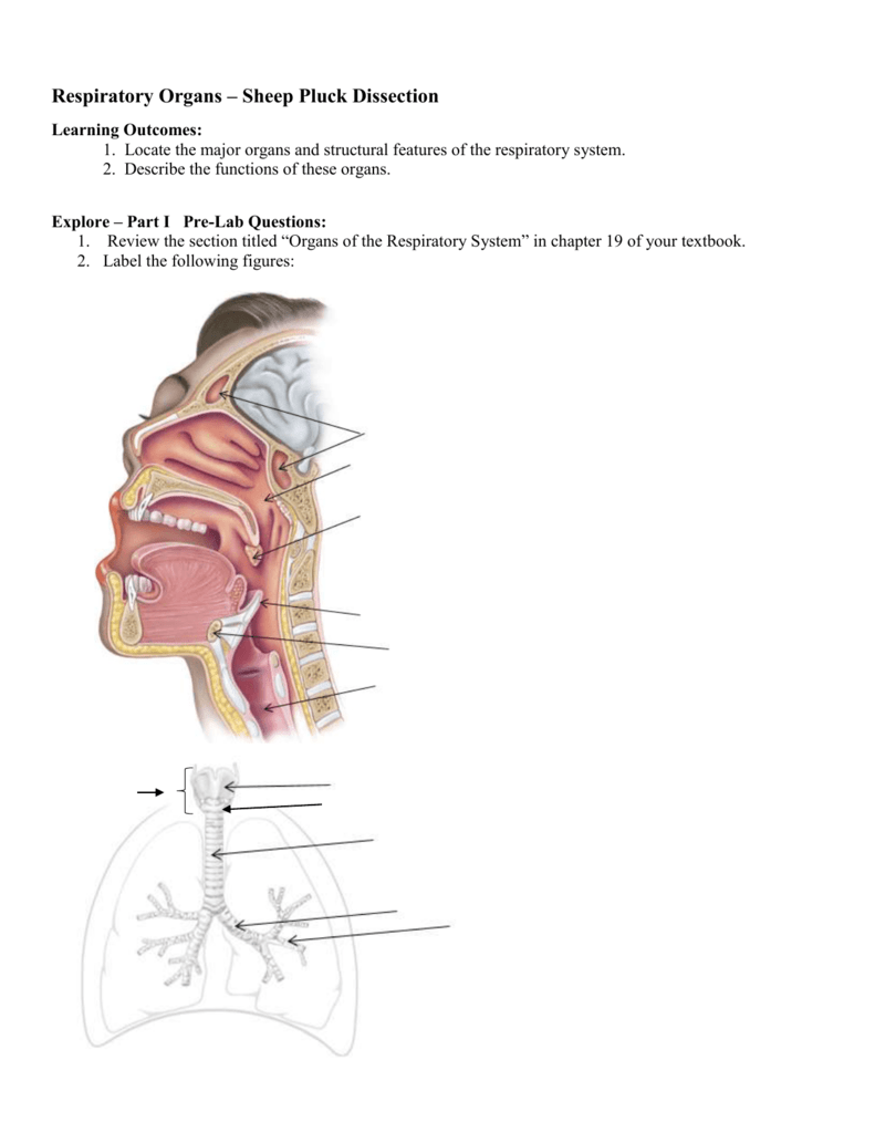
Click for full size pdf. External view of the sheep heart with probes showing the opening to the vena cava and pulmonary vein. Prepare disinfectant solution according to manufacturer s instructions. Cut a piece of the lung tissue out and you should be able to see the tubes branching out. Include a depiction of the trachea and primary bronchus.
Source:
These are called semi lunar valves and prevent blood from flowing backwards from the arteries. It also demonstrates basic skills using a scalpel this video was produce. This should be a clear lump which feels soft on the outside and hard in the middle. Place disinfectant in a container ready for instruments to be placed at the end of the dissection. External view of the sheep heart with probes showing the opening to the vena cava and pulmonary vein.
 Source: studylib.net
Click for full size pdf. Describe the color shape size and feel of the specimen. Place disinfectant in a container ready for instruments to be placed at the end of the dissection. Shows the completed dissection of the sheep heart. When handling the sheep s pluck make sure to wear disposable gloves.
 Source: homesciencetools.com
Into the superior vena cava and make an incision down through the wall of the right atrium and ventricle as shown by the dotted line in the external heart picture. Shows the completed dissection of the sheep heart. Include a depiction of the trachea and primary bronchus. Remember to draw your illustrations to scale. Take care to clearly illustrate and label the four lobes.
 Source: coursehero.com
Aprons and goggles are also available for additional protection. This resource was funded by the gatsby charitable foundation and. It also demonstrates basic skills using a scalpel this video was produce. As seen in the picture to the left. If any blood is associated with the lungs or pluck rinse them in cold running water.
Source:
Cut a piece of the lung tissue out and you should be able to see the tubes branching out. Place disinfectant in a container ready for instruments to be placed at the end of the dissection. Aprons and goggles are also available for additional protection. Remember to draw your illustrations to scale. Distribute the instruments to students.
 Source: coursehero.com
This dissection focuses on the function of the breathing system and a description is provided in the teachers notes of how to inflate one of the lobes in the lungs via an incision made in the trachea. Draw a picture of the right lung of the sheep. Cut a piece of the lung tissue out and you should be able to see the tubes branching out. Describe the color shape size and feel of the specimen. We observed a lung and were of course able to identify the different parts of the.
 Source: coursehero.com
This should be a clear lump which feels soft on the outside and hard in the middle. This dissection focuses on the function of the breathing system and a description is provided in the teachers notes of how to inflate one of the lobes in the lungs via an incision made in the trachea. External view of the sheep heart with probes showing the opening to the vena cava and pulmonary vein. Include a depiction of the trachea and primary bronchus. Aprons and goggles are also available for additional protection.
If you find this site adventageous, please support us by sharing this posts to your own social media accounts like Facebook, Instagram and so on or you can also bookmark this blog page with the title sheep pluck dissection instructions by using Ctrl + D for devices a laptop with a Windows operating system or Command + D for laptops with an Apple operating system. If you use a smartphone, you can also use the drawer menu of the browser you are using. Whether it’s a Windows, Mac, iOS or Android operating system, you will still be able to bookmark this website.


