Staining blood smear
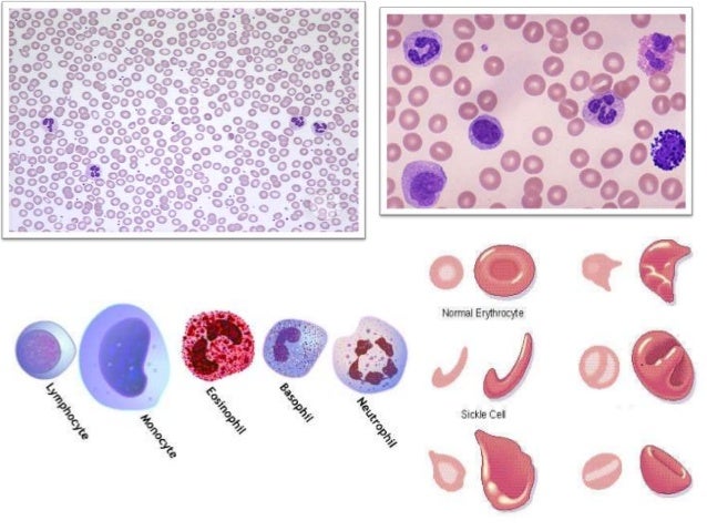
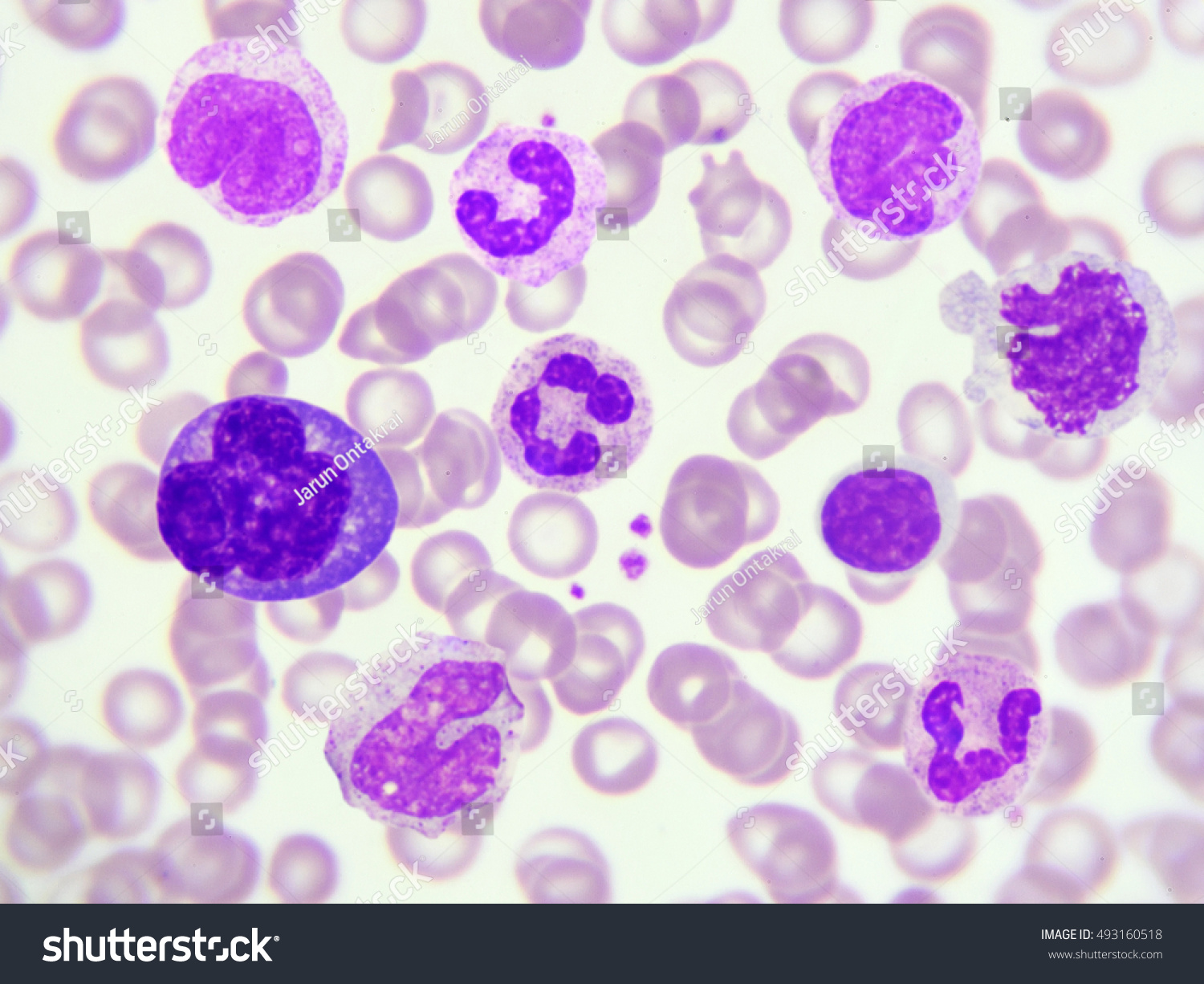
Staining Blood Smear. A blood film or peripheral blood smear is a thin layer of blood smeared on a microscope slide and then stained in such a way to allow the various blood cells to be examined microscopically. The slide is covered with leishman stain for 2 mins. After 2 mins it is diluted with double the volume of buffer water. Staining of blood film.
 Preparation Of Blood Smear From slideshare.net
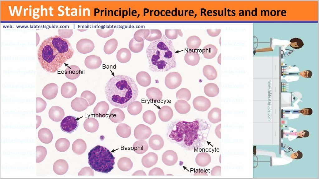
After the blood smear is treated for the proper length of time with wright s stain neutral distilled water is used for diluting the stain. Selection of a spreader. On adding buffer water a metallic shin will be formed if the stain is dry. After 2 mins it is diluted with double the volume of buffer water. Wright s stain is a type of romanowsky stain which is commonly used in hematology laboratory for the routine staining of peripheral blood smears. Example of properly prepared thick and thin film blood smears.
Take one slide a spreader which has smooth edge.
Selection of a spreader. The slide is covered with leishman stain for 2 mins. Staining of blood film. After the slide has been treated with neutral distilled water until the smear becomes pinkish it is then treated with pure absolute methyl alcohol which destains the plasma. Making and staining a blood smear a well made blood smear is a beauty to behold and likely to yield interesting and significant information for a research project. Wright s stain is a type of romanowsky stain which is commonly used in hematology laboratory for the routine staining of peripheral blood smears.
 Source: labtestsguide.com
A preparation of blood smear. It should be done by careful look on the narrow edge of the slide or by moving a thumb smoothly on its edge. Making and staining a blood smear a well made blood smear is a beauty to behold and likely to yield interesting and significant information for a research project. Take one slide a spreader which has smooth edge. This much time is required for fixation.
 Source: shutterstock.com
Selection of a spreader. A blood film or peripheral blood smear is a thin layer of blood smeared on a microscope slide and then stained in such a way to allow the various blood cells to be examined microscopically. It should be done by careful look on the narrow edge of the slide or by moving a thumb smoothly on its edge. The extra time and care taken during the field season will be rewarded later when the smears must be scanned and parasites identified and counted. After 2 mins it is diluted with double the volume of buffer water.
 Source: amazon.com
The smear is covered with stain for approximately ten minutes then diluted with water and allowed an additional ten minutes for the cells to properly stain. The extra time and care taken during the field season will be rewarded later when the smears must be scanned and parasites identified and counted. The smear is covered with stain for approximately ten minutes then diluted with water and allowed an additional ten minutes for the cells to properly stain. After the blood smear is treated for the proper length of time with wright s stain neutral distilled water is used for diluting the stain. It should be done by careful look on the narrow edge of the slide or by moving a thumb smoothly on its edge.
 Source: en.wikipedia.org
After 2 mins it is diluted with double the volume of buffer water. A preparation of blood smear. It is also used for staining bone marrow aspirates urine samples and to demonstrate malarial parasites in blood smears. Following the stain application the. A blood film or peripheral blood smear is a thin layer of blood smeared on a microscope slide and then stained in such a way to allow the various blood cells to be examined microscopically.
 Source: m.youtube.com
Wright s stain is named for james homer wright who devised the stain in 1902 based on a modification of romanowsky stain. A blood film or peripheral blood smear is a thin layer of blood smeared on a microscope slide and then stained in such a way to allow the various blood cells to be examined microscopically. A preparation of blood smear. Blood smear is prepared stained with leishman s stain and cells are identified under oil immersion lens. The slide is covered with leishman stain for 2 mins.
 Source: sigmaaldrich.com
Selection of a spreader. It should be done by careful look on the narrow edge of the slide or by moving a thumb smoothly on its edge. Wright s stain is a type of romanowsky stain which is commonly used in hematology laboratory for the routine staining of peripheral blood smears. Following the stain application the. Blood smear is prepared stained with leishman s stain and cells are identified under oil immersion lens.
 Source: paramedicsworld.com
The smear is covered with stain for approximately ten minutes then diluted with water and allowed an additional ten minutes for the cells to properly stain. Example of properly prepared thick and thin film blood smears. On adding buffer water a metallic shin will be formed if the stain is dry. Selection of a spreader. It is also used for staining bone marrow aspirates urine samples and to demonstrate malarial parasites in blood smears.
 Source: researchgate.net
The extra time and care taken during the field season will be rewarded later when the smears must be scanned and parasites identified and counted. Example of properly prepared thick and thin film blood smears. A preparation of blood smear. Selection of a spreader. This much time is required for fixation.
 Source: en.wikipedia.org
This much time is required for fixation. A preparation of blood smear. Example of properly prepared thick and thin film blood smears. It is also used for staining bone marrow aspirates urine samples and to demonstrate malarial parasites in blood smears. On adding buffer water a metallic shin will be formed if the stain is dry.
 Source: quizlet.com
Following the stain application the. Wright s stain is named for james homer wright who devised the stain in 1902 based on a modification of romanowsky stain. Blood smear is prepared stained with leishman s stain and cells are identified under oil immersion lens. On adding buffer water a metallic shin will be formed if the stain is dry. After the slide has been treated with neutral distilled water until the smear becomes pinkish it is then treated with pure absolute methyl alcohol which destains the plasma.
 Source: slideshare.net
The extra time and care taken during the field season will be rewarded later when the smears must be scanned and parasites identified and counted. Take one slide a spreader which has smooth edge. A blood film or peripheral blood smear is a thin layer of blood smeared on a microscope slide and then stained in such a way to allow the various blood cells to be examined microscopically. Wright s stain is named for james homer wright who devised the stain in 1902 based on a modification of romanowsky stain. The slide is covered with leishman stain for 2 mins.
 Source: paramedicsworld.com
A blood film or peripheral blood smear is a thin layer of blood smeared on a microscope slide and then stained in such a way to allow the various blood cells to be examined microscopically. Take one slide a spreader which has smooth edge. Blood smear is prepared stained with leishman s stain and cells are identified under oil immersion lens. Example of properly prepared thick and thin film blood smears. It should be done by careful look on the narrow edge of the slide or by moving a thumb smoothly on its edge.
 Source: researchgate.net
Wright s stain is named for james homer wright who devised the stain in 1902 based on a modification of romanowsky stain. Blood smear is prepared stained with leishman s stain and cells are identified under oil immersion lens. The extra time and care taken during the field season will be rewarded later when the smears must be scanned and parasites identified and counted. After 2 mins it is diluted with double the volume of buffer water. The slide is covered with leishman stain for 2 mins.
 Source: quizlet.com
After the blood smear is treated for the proper length of time with wright s stain neutral distilled water is used for diluting the stain. Selection of a spreader. Take one slide a spreader which has smooth edge. A preparation of blood smear. Wright s stain is a type of romanowsky stain which is commonly used in hematology laboratory for the routine staining of peripheral blood smears.
 Source: sigmaaldrich.com
Wright s stain is named for james homer wright who devised the stain in 1902 based on a modification of romanowsky stain. Example of properly prepared thick and thin film blood smears. The slide is covered with leishman stain for 2 mins. After the slide has been treated with neutral distilled water until the smear becomes pinkish it is then treated with pure absolute methyl alcohol which destains the plasma. The extra time and care taken during the field season will be rewarded later when the smears must be scanned and parasites identified and counted.
If you find this site convienient, please support us by sharing this posts to your favorite social media accounts like Facebook, Instagram and so on or you can also bookmark this blog page with the title staining blood smear by using Ctrl + D for devices a laptop with a Windows operating system or Command + D for laptops with an Apple operating system. If you use a smartphone, you can also use the drawer menu of the browser you are using. Whether it’s a Windows, Mac, iOS or Android operating system, you will still be able to bookmark this website.


