The cow eye dissection lab
The Cow Eye Dissection Lab. You ll get see how a cow s eye is like a human eye as you closely investigate parts from the iris and lens to the colorful choroid layer. As you get closer to the actual eyeball you may notice muscles that are attached directly to the sclera and along the optic nerve. A preserved cow eye a full color photographic eye dissection guide and the dissection tools lab equipment you ll need. Cats like cows have a tapetum.
Http Ctitus Costello Weebly Com Uploads 5 9 1 1 59117619 Lab Virtualeyedissect Pdf From
A cat s eye seems to glow because the cat s tapetum is reflecting light. You should be able to find the sclera or the whites of the eye. The eye most likely has a thick covering of fat and muscle tissue. Leave the optic nerve attached. My hypothesis was that as i was examining and labeling theat the smaller less defined features would be harder to locate. Cut the eyeball in half.
For this experiment i took the cow eye and removed all fat and muscle around the eye then cut it open and.
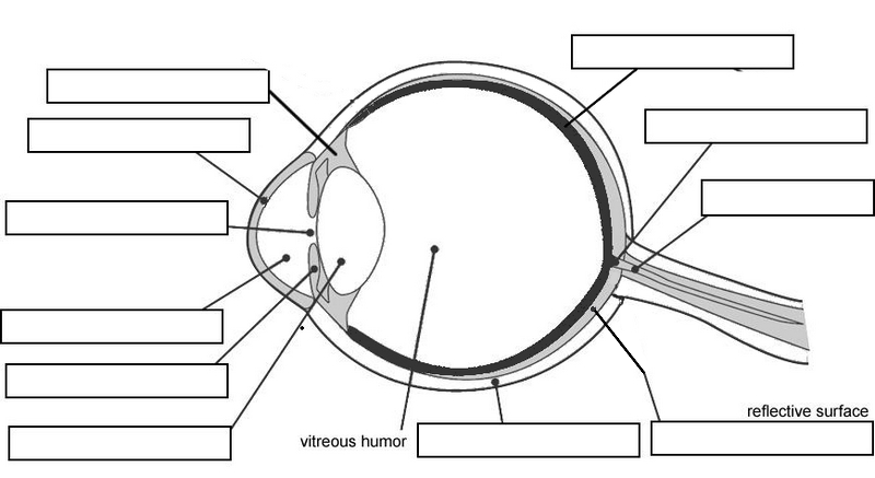
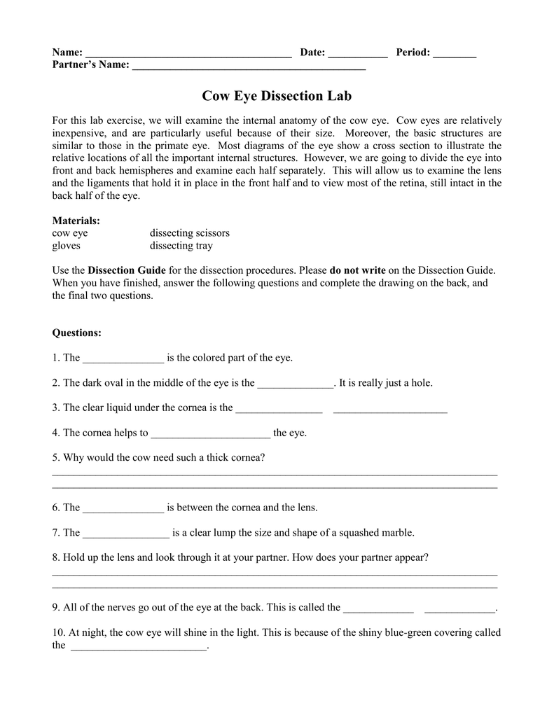
This is the tapetum. Cow eye and sheep s brain dissection abstract the purpose of the experiment was to give us hands on experience with the structures of the eye and of the brain. When the cow was alive the cornea was clear. Cow eye dissection worksheet 1. Cut the eyeball in half. Locate the covering over the front of the eye the cornea.
 Source: biologycorner.com
As you get closer to the actual eyeball you may notice muscles that are attached directly to the sclera and along the optic nerve. During this activity you will dissect a cow eye. As you get closer to the actual eyeball you may notice muscles that are attached directly to the sclera and along the optic nerve. Learn how to dissect a cow s eye in your classroom. Cow eye dissection worksheet 1.
 Source: promotiontablecovers.blogspot.com
Follow step 1 in the cow s dissection guide. These are the extrinsic muscles that allow a cow to move its eye up and down and from side to side. View cow eye dissection lab pdf from zoo 102 at saint mary s college of california. A 22 broad blade scalpel scissors and a sturdy disposable dissecting tray. You ll get see how a cow s eye is like a human eye as you closely investigate parts from the iris and lens to the colorful choroid layer.
 Source: studylib.net
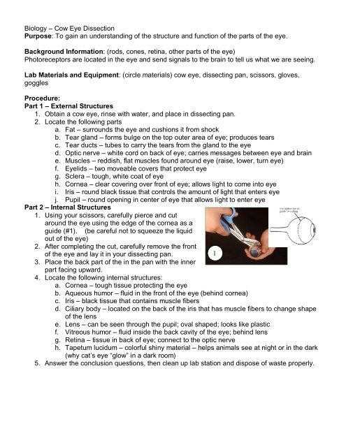
Tapetum mid layer of the eye and contains reflective pigments that helps the eye see better at night. Check out our online cow eye dissection project with pictures. Remove excess tissue from the eye such as the fat muscle eye lids etc. Carefully cut away the fat and the muscle. Learn how to dissect a cow s eye in your classroom.
 Source: studylib.net
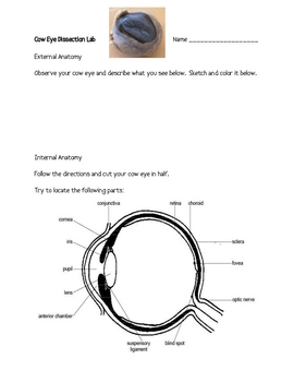
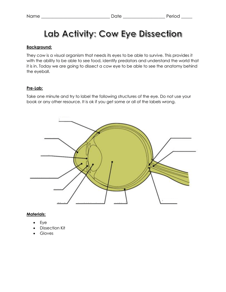
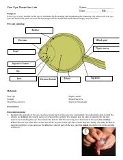
S clera a tough white outermost covering of the eye. Carefully cut away the fat and the muscle. This is the tapetum. When the cow was alive the cornea was clear. L ens clear flexible structure that focuses the light on the retina.
Source:
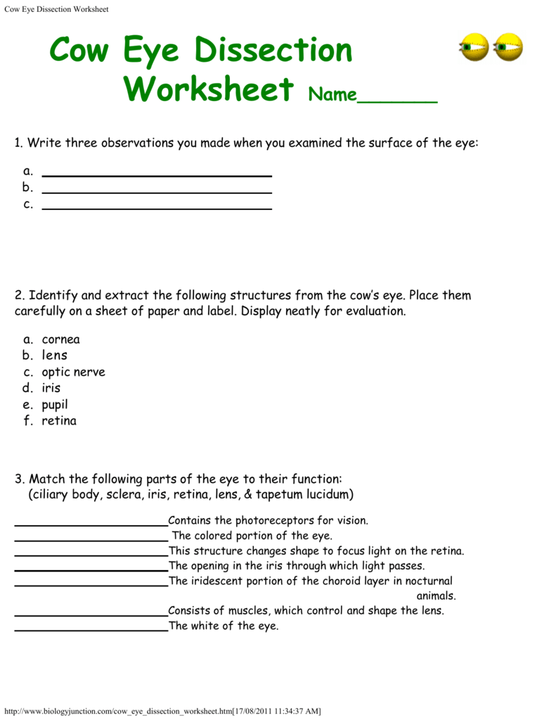
Materials preserved cow eye scalpel or scissors forceps dissection tray gloves safety glasses lab apron 1. Have you ever seen a cat s eyes shining in the headlights of a car. A step by step hints and tips a cow eye primer and a glossary of terms. As you get closer to the actual eyeball you may notice muscles that are attached directly to the sclera and along the optic nerve. Cow eye dissection worksheet 1.
 Source: yumpu.com
L ens clear flexible structure that focuses the light on the retina. This is the tapetum. Cow eye dissection worksheet 1. These are the extrinsic muscles that allow a cow to move its eye up and down and from side to side. A preserved cow eye a full color photographic eye dissection guide and the dissection tools lab equipment you ll need.
 Source: pinterest.com
You will observe several important features of the eye and develop your understanding of how each part functions to make vision possible. Cut the eyeball in half. You ll get see how a cow s eye is like a human eye as you closely investigate parts from the iris and lens to the colorful choroid layer. Tapetum mid layer of the eye and contains reflective pigments that helps the eye see better at night. These are the extrinsic muscles that allow a cow to move its eye up and down and from side to side.
 Source: studylib.net
Materials preserved cow eye scalpel or scissors forceps dissection tray gloves safety glasses lab apron 1. As you get closer to the actual eyeball you may notice muscles that are attached directly to the sclera and along the optic nerve. Locate the covering over the front of the eye the cornea. View cow eye dissection lab pdf from zoo 102 at saint mary s college of california. Place the cow eye on a dissecting tray.
 Source: nematodejaqw.de.tl
Examine the outside of the eye. Need a preview before you commit. Place the cow eye on a dissecting tray. View cow eye dissection lab pdf from zoo 102 at saint mary s college of california. Take pictures of each step of the eye process.
Source: biologycorner.com
Cut the eyeball in half. Obtain a cow eye dissection styrofoam tray gloves and surgical tools. Materials preserved cow eye scalpel or scissors forceps dissection tray gloves safety glasses lab apron 1. Follow step 1 in the cow s dissection guide. When the cow was alive the cornea was clear.
 Source: pinterest.com
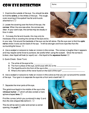
With a sharp point poke a whole on the side of the eye to relieve the pressure in the eyeball so you wont get squirted with liquid when cutting open the eyeball. Materials preserved cow eye scalpel or scissors forceps dissection tray gloves safety glasses lab apron 1. Leave the optic nerve attached. The eye most likely has a thick covering of fat and muscle tissue. As you get closer to the actual eyeball you may notice muscles that are attached directly to the sclera and along the optic nerve.
 Source: studylib.net
During this activity you will dissect a cow eye. Cow s eye dissection page 7 under the retina the back of the eye is covered with shiny blue green stuff. These are the extrinsic muscles that allow a cow to move its eye up and down and from side to side. Examine the outside of the eye. Follow step 1 in the cow s dissection guide.
 Source: biologycorner.com
Cow s eye dissection page 7 under the retina the back of the eye is covered with shiny blue green stuff. Cats like cows have a tapetum. Cut the eyeball in half. Cow s eye dissection page 7 under the retina the back of the eye is covered with shiny blue green stuff. Carefully cut away the fat and the muscle.
 Source: coursehero.com
This is the tapetum. Cow eye dissection worksheet 1. Keep cutting close to the sclera separating the membrane that attaches the muscle to it. C horoid layer mid layer of the eye and contains dark pigments that helps nourish the retina. A cat s eye seems to glow because the cat s tapetum is reflecting light.
 Source: coursehero.com
These are the extrinsic muscles that allow a cow to move its eye up and down and from side to side. You ll get see how a cow s eye is like a human eye as you closely investigate parts from the iris and lens to the colorful choroid layer. Have you ever seen a cat s eyes shining in the headlights of a car. C horoid layer mid layer of the eye and contains dark pigments that helps nourish the retina. A cat s eye seems to glow because the cat s tapetum is reflecting light.
If you find this site value, please support us by sharing this posts to your preference social media accounts like Facebook, Instagram and so on or you can also bookmark this blog page with the title the cow eye dissection lab by using Ctrl + D for devices a laptop with a Windows operating system or Command + D for laptops with an Apple operating system. If you use a smartphone, you can also use the drawer menu of the browser you are using. Whether it’s a Windows, Mac, iOS or Android operating system, you will still be able to bookmark this website.


