Thoracic cavity of a fetal pig
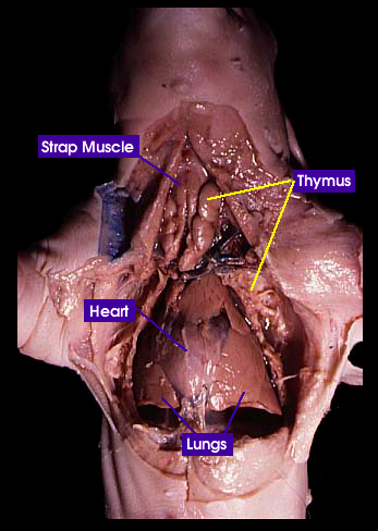
Thoracic Cavity Of A Fetal Pig. You will need to cut through the pig s sternum and expose the chest cavity thoracic cavity. These muscles when contracted increase the thoracic cavity size during inhalation to allow for the air to rush in. Two spongy like surround the heart lungs weren t used by fetus so they contain no air. A similar cut is made on the other side.

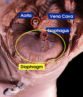
The abdominal cavity is separated from the thoracic cavity by the diaphragm. It is a dome shaped sheet of muscle and tendon that also serve in respiration and the breathing process. Try using search on phones and tablets. These muscles when contracted increase the thoracic cavity size during inhalation to allow for the air to rush in. A cut is made on the side of the animal from the point just posterior to the diaphragm dorsally. Self quiz video link at the end of part 4.
Trachea large tube lies anterior to the lungs.
The thoracic cavity and the abdominal cavity are separated by the diaphragm. Opening the abdominal cavity. A cut is made on the side of the animal from the point just posterior to the diaphragm dorsally. Note the many membranes lining the coelom and holding the organs in place. Trachea large tube lies anterior to the lungs. Dissection of the thoracic cavity.
 Source: commons.wikimedia.org
In mammals the coelom is divided into two main cavities. A cut is made on the side of the animal from the point just posterior to the diaphragm dorsally. Try using search on phones and tablets. To study the thoracic cavity and urogenital system of the fetal pig. The action of breathing is a muscular operation the muscles involved are.
 Source: quizlet.com
Two spongy like surround the heart lungs weren t used by fetus so they contain no air. Find the diaphragm again. Note the many membranes lining the coelom and holding the organs in place. These muscles when contracted increase the thoracic cavity size during inhalation to allow for the air to rush in. You will need to cut through the pig s sternum and expose the chest cavity thoracic cavity.
 Source: canyons.edu
Dissection of the thoracic cavity. Self quiz video link at the end of part 4. A cut is made on the side of the animal from the point just posterior to the diaphragm dorsally. These muscles when contracted increase the thoracic cavity size during inhalation to allow for the air to rush in. Find the diaphragm again.

Sac surrounding the heart and the diaphragm from the body wall. In mammals the coelom is divided into two main cavities. Sac surrounding the heart and the diaphragm from the body wall. Above the diaphragm center of chest is the heart. The abdominal cavity is separated from the thoracic cavity by the diaphragm.
 Source: whitman.edu
The diaphragm which separates the thoracic from the abdominal cavity and the intercostal muscles found between the ribs bone. Opening the abdominal cavity. These two cuts will enable you to spread open the abdominal cavity. Part 2 of 4 click the annotation for the next part. You will need to cut through the pig s sternum and expose the chest cavity thoracic cavity.
 Source: cals.ncsu.edu
Self quiz video link at the end of part 4. To study the thoracic cavity and urogenital system of the fetal pig. Navigation best viewed on larger screens. The thoracic cavity which contains the lungs and the abdominal cavity which contains the digestive system. Sac surrounding the heart and the diaphragm from the body wall.
 Source: canyons.edu
Part 2 of 4 click the annotation for the next part. Navigation best viewed on larger screens. In mammals the coelom is divided into two main cavities. You will need to cut through the pig s sternum and expose the chest cavity thoracic cavity. It is a dome shaped sheet of muscle and tendon that also serve in respiration and the breathing process.
 Source: people.hsc.edu
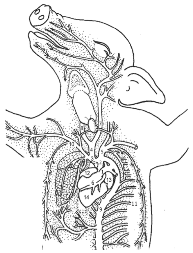
Navigation best viewed on larger screens. Home academics biology resources biology lab 107 resources histology anatomy of fetal pig thoracic cavity histology anatomy of fetal pig jugular veins jugular veins the jugular veins are veins that bring deoxygenated blood from the head back to the heart via the superior vena cava. Remember that the diaphragm separates the abdominal cavity from the thoracic cavity and it aids in breathing. In mammals the coelom is divided into two main cavities. Above the diaphragm center of chest is the heart.

The thoracic cavity and the abdominal cavity are separated by the diaphragm. The diaphragm which separates the thoracic from the abdominal cavity and the intercostal muscles found between the ribs bone. Find the diaphragm again. Remember that the diaphragm separates the abdominal cavity from the thoracic cavity and it aids in breathing. Navigation best viewed on larger screens.
 Source: courses.lumenlearning.com
You will need to cut through the pig s sternum and expose the chest cavity thoracic cavity. The thoracic cavity and the abdominal cavity are separated by the diaphragm. It is a dome shaped sheet of muscle and tendon that also serve in respiration and the breathing process. Above the diaphragm center of chest is the heart. Remember that the diaphragm separates the abdominal cavity from the thoracic cavity and it aids in breathing.
 Source: bio.libretexts.org
Navigation best viewed on larger screens. Above the diaphragm center of chest is the heart. Opening the abdominal cavity. Part 2 of 4 click the annotation for the next part. Try using search on phones and tablets.
 Source: whitman.edu
To study the thoracic cavity and urogenital system of the fetal pig. The thoracic cavity and the abdominal cavity are separated by the diaphragm. Mouth and neck region. Try using search on phones and tablets. The thoracic cavity which contains the lungs and the abdominal cavity which contains the digestive system.
 Source: pinterest.com
Opening the abdominal cavity. Note the many membranes lining the coelom and holding the organs in place. Navigation best viewed on larger screens. Part 2 of 4 click the annotation for the next part. These two cuts will enable you to spread open the abdominal cavity.
 Source: courses.lumenlearning.com
Home academics biology resources biology lab 107 resources histology anatomy of fetal pig thoracic cavity histology anatomy of fetal pig jugular veins jugular veins the jugular veins are veins that bring deoxygenated blood from the head back to the heart via the superior vena cava. In mammals the coelom is divided into two main cavities. You will need to cut through the pig s sternum and expose the chest cavity thoracic cavity. Part 2 of 4 click the annotation for the next part. To study the thoracic cavity and urogenital system of the fetal pig.

Home academics biology resources biology lab 107 resources histology anatomy of fetal pig thoracic cavity histology anatomy of fetal pig jugular veins jugular veins the jugular veins are veins that bring deoxygenated blood from the head back to the heart via the superior vena cava. A similar cut is made on the other side. Two spongy like surround the heart lungs weren t used by fetus so they contain no air. A cut is made on the side of the animal from the point just posterior to the diaphragm dorsally. You will need to cut through the pig s sternum and expose the chest cavity thoracic cavity.
If you find this site convienient, please support us by sharing this posts to your preference social media accounts like Facebook, Instagram and so on or you can also save this blog page with the title thoracic cavity of a fetal pig by using Ctrl + D for devices a laptop with a Windows operating system or Command + D for laptops with an Apple operating system. If you use a smartphone, you can also use the drawer menu of the browser you are using. Whether it’s a Windows, Mac, iOS or Android operating system, you will still be able to bookmark this website.


